

The growth of endometrial tissue outside the uterus can lead to significant pain and infertility in women. Endometriosis causes scarring and implants on the ovaries, fallopian tubes, outer uterine wall, vaginal wall, intestines and bladder. These implants can vary from microscopic sizes to large cysts, known as endometriomas.
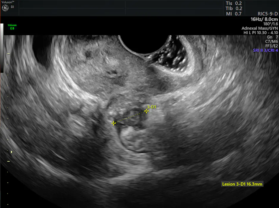
lesion on endometriosis ultrasound
Implants that penetrate the retroperitoneal space to a depth of 5 mm or more are classified as deep infiltrating endometriosis (DIE). DIE can involve the pouch of Douglas, the rectovaginal septum, intestines, anterior pouch and the uterosacral ligaments.
Diagnosing endometriosis is difficult to accomplish by physical examination only and requires a reliable diagnostic imaging exam. Additionally, preoperative imaging is crucial for identifying the different locations of deep endometriosis because in certain sites, such as the intestine or bladder, the surgery is particularly difficult and carries greater risk.
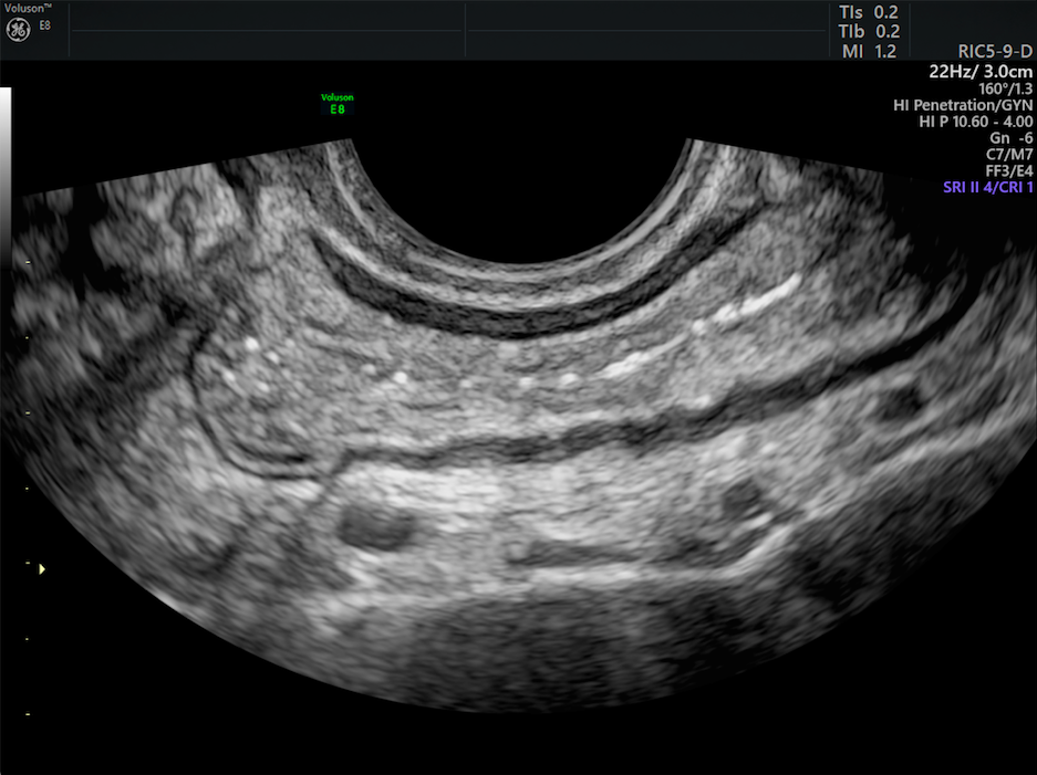
Normal bowel wall on endometriosis ultrasound
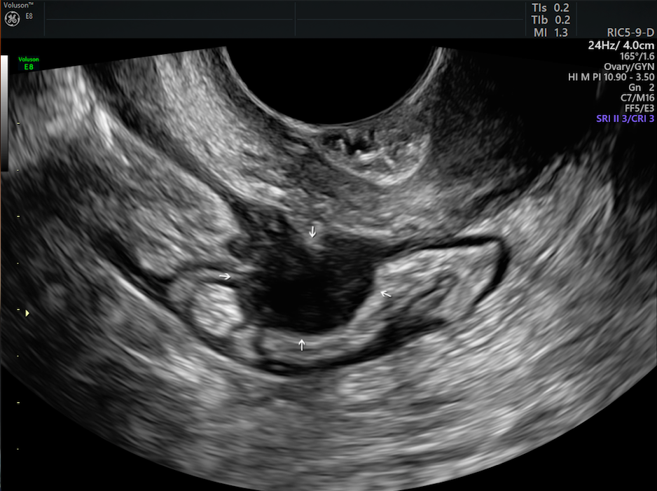
Deep Infiltrating Endometriosis in bowel wall
Laparoscopy is the standard method to diagnose pelvic endometriosis. For non-invasive options, however, endometriosis ultrasound and MRI are the two most commonly used imaging methods.
Diagnosing Endometriosis With MRI
MRI is helpful in determining the extent of deep infiltrating endometriosis, especially when laparoscopic inspection is limited by adhesions, as reported by a study in The Egyptian Journal of Radiology and Nuclear Medicine. It also offers better differentiation between endometriomas and hemorrhagic cysts, which can appear similar when using ultrasound.
MRI can help direct surgical approaches for patients with suspected endometriosis, especially for DIE and other uncommon areas of occurrence. Also, the assessment of ectopic endometriosis in extrapelvic locations — most commonly within muscle, cutaneous tissue and the thorax — is better addressed by MRI.
Diagnosing Endometriosis on Transvaginal Ultrasound in 3D
Ultrasound is very effective in the identification of large endometriosis lesions. As reported in the same study in The Egyptian Journal of Radiology and Nuclear Medicine, transvaginal ultrasound can help diagnose small endometriomas, bladder lesions and deep nodules, including those in the rectovaginal septum. Ultrasound also offers Doppler blood flow mapping of the pelvis without the need for contrast.
Compared to 2D ultrasound, 3D ultrasound is much more accurate in diagnosing posterior locations of deep endometriosis without intestinal involvement, such as the uterosacral ligaments and vaginal and rectovaginal areas, according to a study in Human Reproduction. 3D volume ultrasound imaging, with its automated acquisition, can generate hundreds of images and be used to reconstruct any view in any orientation. This imaging technology also has good reproducibility even when operated by a practitioner with less expertise.
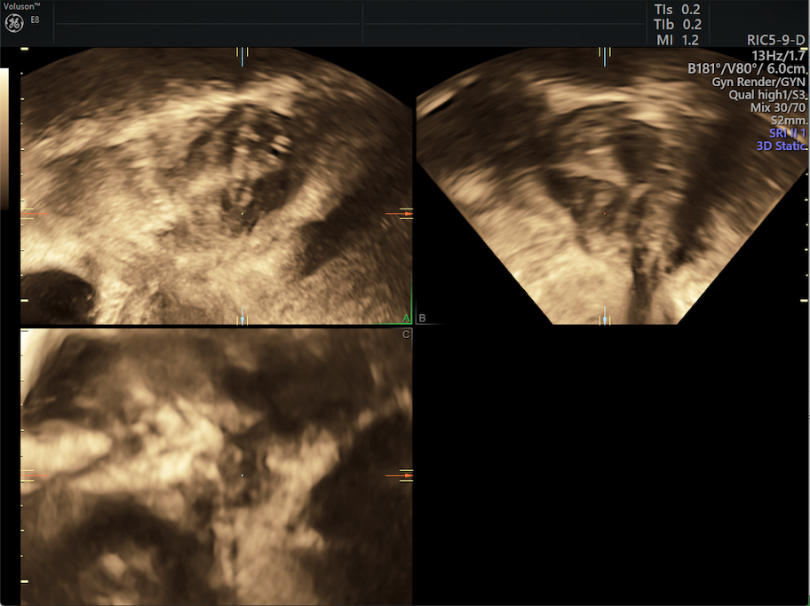
3D images of deep infiltrating endometriosis on transvaginal ultrasound.
Transvaginal ultrasound that is "tenderness-guided" evaluates painful sites with a gentle pressure of the probe, and it has the benefit of being able to look more directly at areas of pain within the pelvis while getting real-time feedback from the patient and assessing the motion of structures.
Determining the Best Non-Invasive Assessment Method
A 2017 meta‐analysis of studies, published by Ultrasound in Obstetrics & Gynecology, compared the use of transvaginal ultrasound and MRI in the assessment of endometriosis, including deep infiltrating endometriosis. The results demonstrated similar diagnostic performance between transvaginal ultrasound and MRI in the detection of endometriomas of the ovaries and fallopian tubes, as well as DIE in the areas of the rectosigmoid, rectovaginal septum and uterosacral ligaments.
However, 3D ultrasound imaging is much more readily accessible, less expensive and less time-consuming than MRI. Real-time endometriosis ultrasound imaging allows for the evaluation of areas of tenderness and the use of the sliding sign for a more thorough assessment of the pelvis. Bowel peristalsis does not affect ultrasound imaging as it does MRI, and many patients express a preference for transvaginal ultrasound because they find it more comfortable than the MRI process. For these reasons, transvaginal ultrasound can be used as a first-line imaging technique when assessing and diagnosing endometriosis.