

When evaluating ovarian mass symptoms in patients, ultrasound is a first-line tool for gynecologists to determine the size, shape, complexity, blood flow and movement of the ovaries. While most masses are not malignant, ovarian cancer is one of the deadliest gynecologic cancers, so it's important to gather as much information as possible during the evaluation. Details and a thorough description of the mass can help you determine next steps and develop a management plan for your patient.
Determining the Risk of Cancer
Before beginning an ovarian mass evaluation, the gynecologist should first consider the patient's risk of cancer. According to RadioGraphics, a woman in the U.S. has a 5 to 10 percent chance of undergoing surgery for a suspected ovarian malignancy during her lifetime. Of those patients, only about 13 to 21 percent will be diagnosed with ovarian cancer.
Further, the risk of ovarian cancer increases with age. In fact, the American Cancer Society reported that half of all ovarian cancers are found in women 63 or older. Women who have carried a pregnancy to term before age 26 or who have been on birth control pills are at a lower risk of cancer.
Looking at the Ovaries on Ultrasound
A transvaginal ultrasound combined with a pelvic exam can provide gynecologists with more information than just an abdominal ultrasound. By gathering data through an ultrasound exam, the physician can create a picture that helps determine whether a mass is cancerous.
When assessing ovarian mass symptoms with ultrasound, gynecologists should consider the following factors:
- Size: Measure both the size of the ovary and the mass. If a mass is 10 cm or larger, according to research published in Obstetrics and Gynecology, the risk of cancer is increased.
- Shape: An irregularly shaped mass is often more suspicious than a smooth, regularly shaped one. A 3D ultrasound can provide a better view of the shape and complexity of the mass in all directions.
- Vascularity: Turning on the color Doppler mode allows you to see the blood flow to ovaries on ultrasound. Increased blood flow could indicate a suspicious mass, in which case you might want to refer the patient to a gynecology oncologist. Cancerous tumors typically contain dilated, saccular and randomly dispersed vessels, according to Gynecology & Obstetrics.
- Density: Determine whether the cyst is solid. According to OBG Management, about 10 percent of solid masses are cancerous. These masses represent the smallest subset of ovarian tumors.
- Fluid: Transvaginal ultrasound can detect cul-de-sac fluid. Fluid is not discovered in the majority of cysts in postmenopausal patients, and fluid in a mass belonging to a postmenopausal woman might be a sign of a tumor.
- Changes over time: Between 53 and 89 percent of functional cysts spontaneously regress, which can be determined by doing a follow-up ultrasound in six to eight weeks, according to OBG Management.
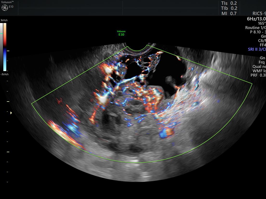
Ultrasound of a serous ovarian cancer FIGO stage IIIB
Image courtesy of Prof. Dirk Timmerman.
When assessing potentially malignant masses, compassionate, high-quality patient care is more crucial than ever. Taking these steps into consideration when examining ovarian masses can help you develop a path forward for your patients as you determine the appropriate course of care.


