In vitro fertilization (IVF) can be a costly and emotional experience for your patients, but a new tool is helping improve their chances of IVF success: time-lapse monitoring of embryos. Time-lapse monitoring is used to help improve embryo selection and increase the chances of transferring a viable embryo for implantation.
Culturing human embryos in vitro requires the maintenance of special conditions to mimic the natural environment. Here's what clinicians need to know about time-lapse monitoring so they can guide patients through this complex process.
Monitoring Embryos: From Snapshots to Moving Pictures
Embryos are kept in incubators in a controlled atmosphere. To evaluate the development of the embryos as they are cultured, the dish of embryos must be removed daily for inspection and assessment of cleavage and morphology.
This peek is simply a one-time snapshot of a dynamic process. Removing the embryos from the ideal culturing conditions stresses them, and there is a limit to the number of assessments that can be performed this way. Instead, using time-lapse monitoring allows for continuous and noninvasive monitoring of embryos.
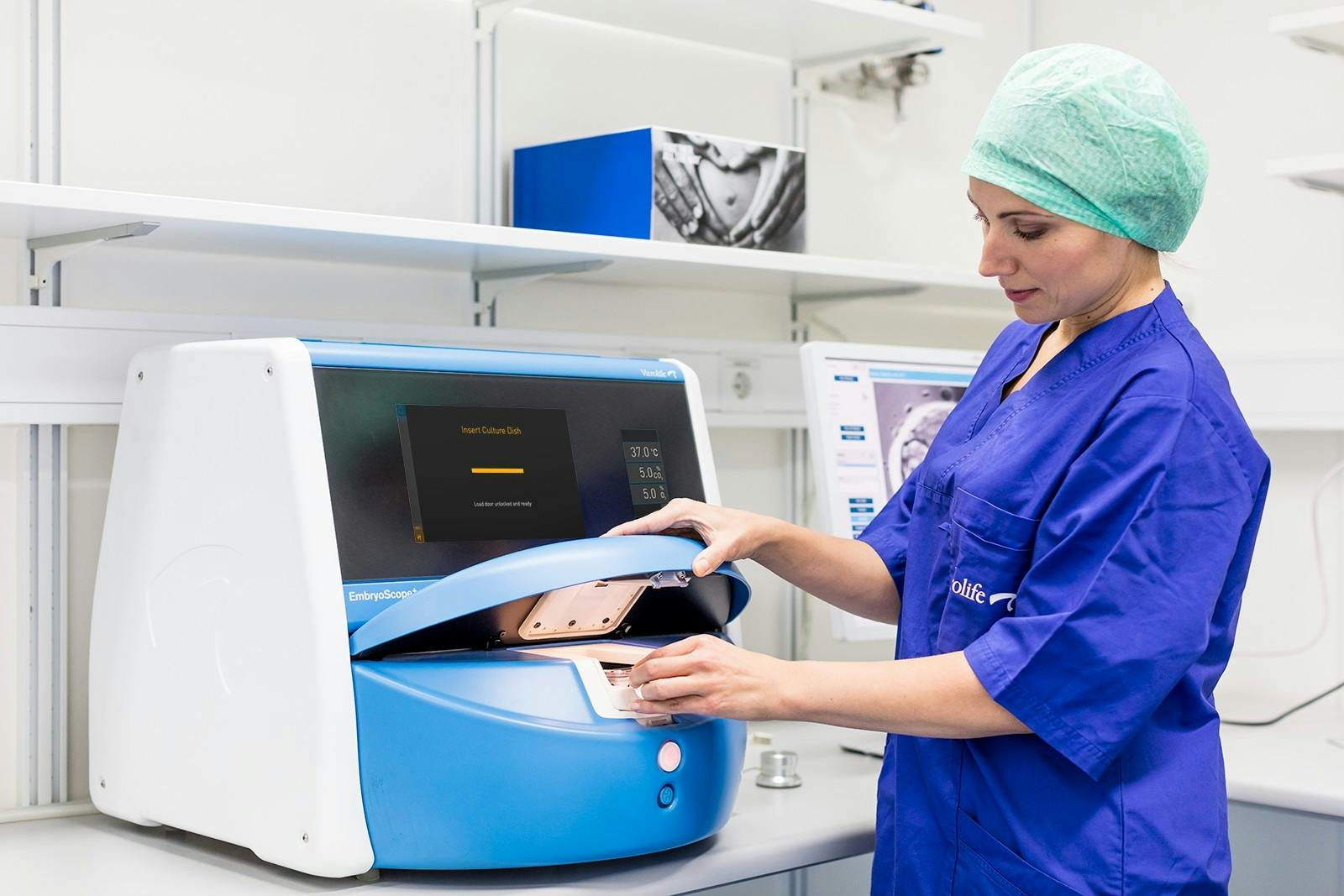
Time-lapse embryo monitoring setups like the EmbryoScope system incorporate a digital inverted microscope into an incubator that provides a stable culture environment for embryos. The microscope takes images of the embryos at set intervals, processes them with custom image acquisition and displays the resulting short time-lapse videos on a computer screen. The system's software allows an embryologist to look at the time-lapse clips of patients' embryos to select the best ones for transfer and freezing.
Observing Embryos for Changes
Observing embryos in this ongoing, dynamic way allows an embryologist to watch more of the important changes that the embryo undergoes, such as cleavage pattern. This visibility is designed to improve a clinician's ability to select the best embryos for transfer. For example, some abnormal cleavages, such as direct cleavage, where one cell divides into more than two daughter cells, can be challenging to identify with standard embryo assessment, according to Vitrolife.
Some studies have connected direct cleavage and other irregular cleavage patterns with lower implantation and pregnancy rates. Using time-lapse monitoring can help identify these patterns and potentially improve embryo selection.
The risk of multiple gestation — and the resulting additional risks to maternal, fetal and neonatal health — could also be lowered with the use of time-lapse embryo monitoring. This process aims to help select embryos with the greatest potential for successful implantation. With closer monitoring, a single embryo transfer could be more confidently performed to eliminate the risk of multiple pregnancies but raise the success rate of pregnancy.
Improving Birth Rates
According to a recent meta-analysis published in Reproductive Biomedicine Online, utilizing time-lapse systems can significantly improve clinical pregnancy rates and live birth rates and reduce early pregnancy loss. These improvements mean a shortened overall time for your patients to achieve a successful pregnancy.
Time-lapse embryo monitoring is a valuable tool that can assist embryologists in selecting embryos with the highest implantation potential. Future studies may reveal even more about its potential to help your patients and your practice.