
Gynecologists no longer have to rely on fuzzy, one-dimensional ultrasound images. New ultrasound technology has leaped forward, allowing practices to replace their 2D ultrasound equipment with superior 3D ultrasound systems.
3D ultrasound produces complete views of the anatomy of the uterus and cervix, providing access to imaging planes not achievable with 2D systems. Gynecologists can use 3D ultrasound to better view and identify abnormalities, verify placement of IUDs and more, which enables them to improve diagnoses, efficiency and patient satisfaction.
The Benefits of New Ultrasound Technology
Ultrasound has been around since the 1950s, although it took a few decades before it became ubiquitous in gynecology. A 1958 paper published in The Lancet described using ultrasound to obtain images of a fetus and gynecological masses for the first time. The technology improved over the following decades, and today 3D ultrasound provides enhanced clarity, faster speeds and more flexibility for gynecology practices.
One of the most significant advantages of 3D ultrasound is that it allows gynecologists to view the coronal plane of the uterus. Combined with enhanced image clarity, 3D ultrasound can improve gynecologists' ability to spot abnormalities that are tiny or otherwise hidden in a 2D image.
3D ultrasound also assists gynecologists with IUD placement. According to Contemporary OB/GYN, the popularity of long-acting reversible contraception (LARC) has increased fivefold in the past decade. Additionally, 8 percent of women ages 15 to 44 now use either an IUD or a contraceptive implant, the Centers for Disease Control and Prevention reported.
A complete view of the coronal plane of the uterus can help gynecologists insert IUDs with greater confidence. The technology can also be beneficial if a physician suspects a patient has a small or large uterus. A 3D ultrasound before IUD insertion can help the doctor with proper placement. Furthermore, if a patient with an IUD experiences pain and/or abnormal bleeding, the physician can use 3D technology to help determine whether the IUD has become displaced.
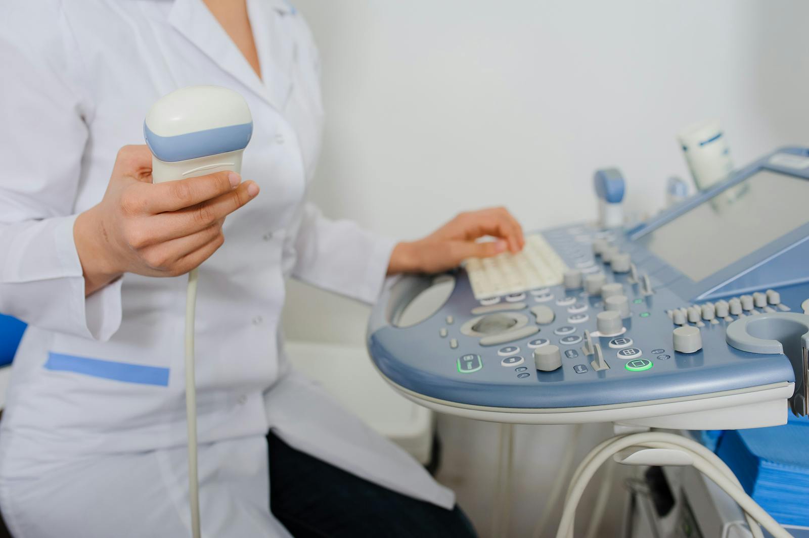
Improvements in Transducer Technology
Over the years, making 3D ultrasound machines easier to use has been a top priority. Attention has been focused on developing portable, convenient systems, making them ideal for independent practices. Transducer technology has also improved, leading to sharper images and lighter, ergonomically designed devices that minimize user fatigue.
Thanks to new 3D ultrasound technology, gynecologists can improve their practices and offer patients efficient, cost-effective options right in their office. Ultimately, 3D ultrasound helps physicians better serve their patients, improving satisfaction and patient-doctor collaboration.


