
A practitioner's first concern upon spotting an ovarian mass on a patient image is often to determine what kind of mass it is and the possible impact on a patient's health. Your patient's first concern, however, is often knowing whether or not that mass means cancer.
Understanding complex ovarian cysts and being able to differentiate them from simple ovarian cysts is paramount for physicians and gynecologists. Being able to explain the risk for ovarian cancer in a straightforward and reassuring way for your concerned patients is equally crucial.
With the help of a growing number of imaging and modeling tools, healthcare professionals are now better equipped than ever to treat ovarian cysts and masses and determine the probability that they will lead to cancer.
Simple vs. Complex Ovarian Cysts
Ovarian cysts are sacs that develop in or on the ovary. There are two main types of cysts: simple ovarian cysts and complex ovarian cysts. Simple ovarian cysts are fluid-filled sacs, and they are fairly common in both premenopausal and postmenopausal women. These sacs don't typically lead to cancer or an increased risk of it, and many simple ovarian cysts will disappear on their own without treatment. In fact, one recent study estimated that fewer than one out of 1,000 women with only a simple ovarian cyst would develop ovarian cancer.
Unlike simple cysts, complex ovarian cysts are solid or irregular masses. Types of complex masses include endometriomas, dermoids and cystadenomas. Although there is a complex ovarian cyst cancer risk, these masses won't necessarily lead to cancer either. The U.S. Department of Health and Human Services estimates that 5 to 10 percent of women have surgery to remove an ovarian cyst, but only 13 to 21 percent of those are cancerous. Gynecologists can use ultrasound technology to distinguish between these different types of ovarian masses.
Rules for Identifying Ovarian Masses
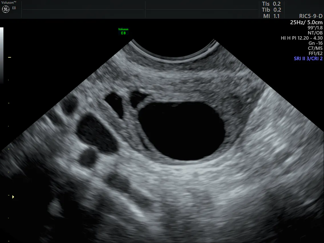
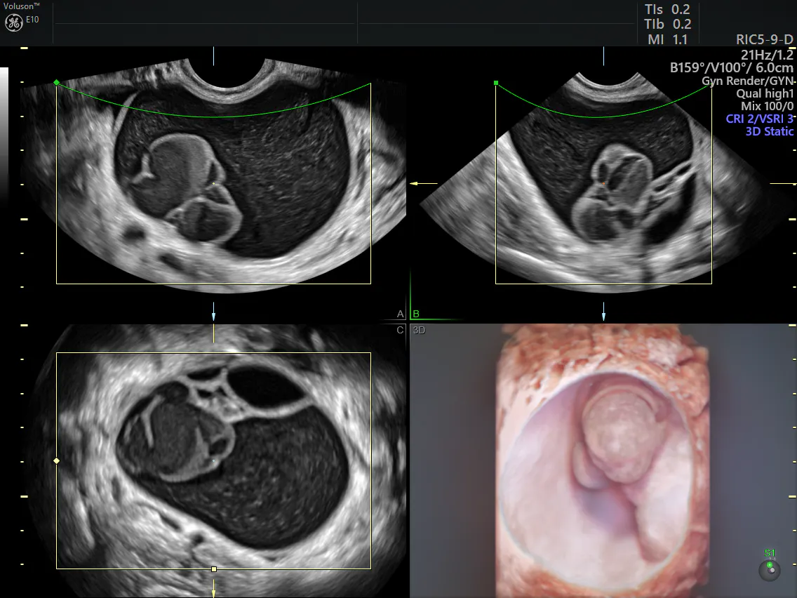
Transvaginal ultrasound, especially 3D ultrasound, can help physicians differentiate between benign simple cysts and potentially cancerous complex ovarian masses. For example, cysts that contain papillary structures, solid areas and increased vascularity are more likely to be malignant. Still, it can be difficult for clinicians to differentiate between some complex and simple ovarian masses.
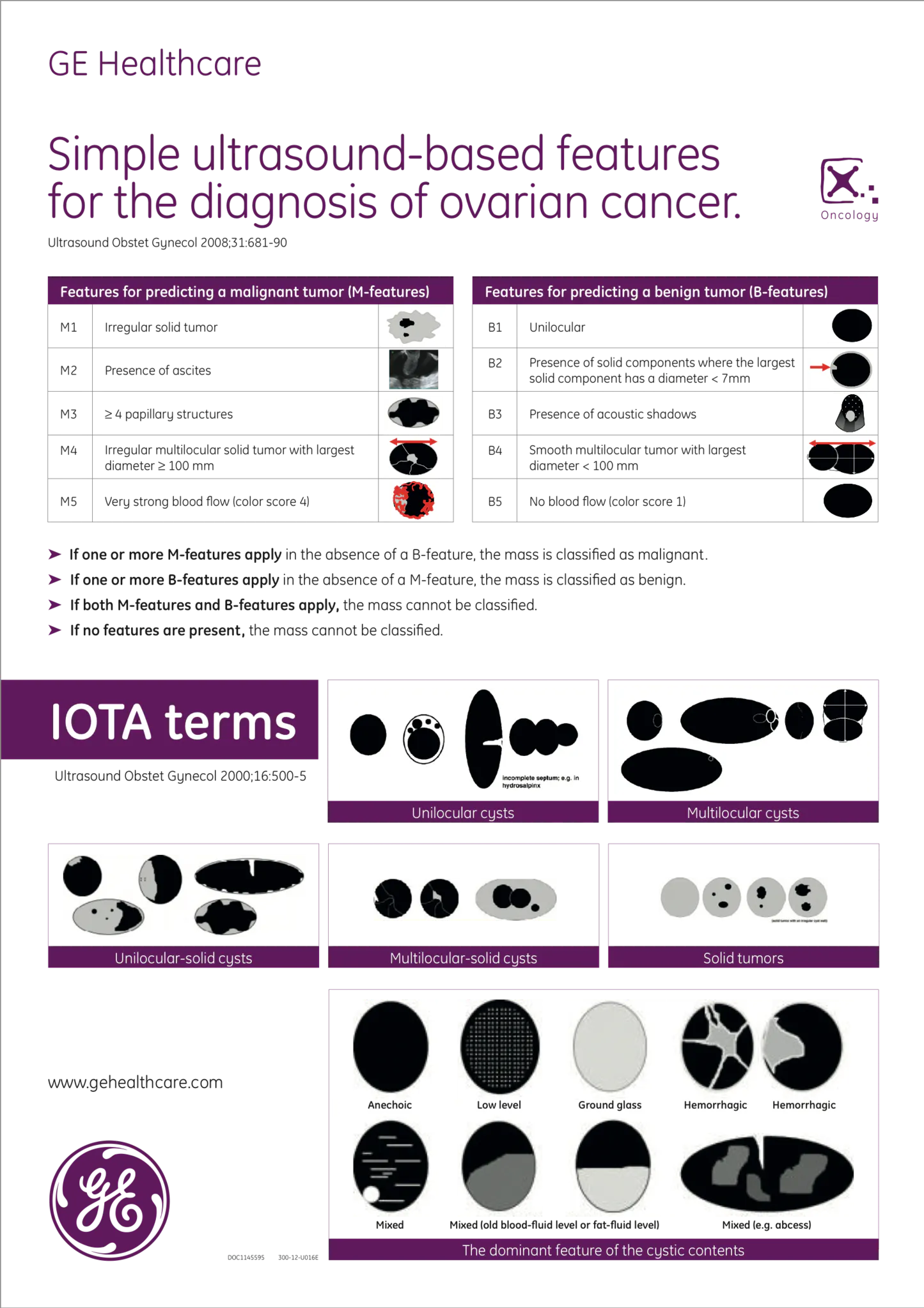
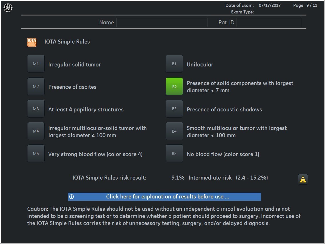
The International Ovarian Tumor Analysis (IOTA) model attempts to clarify these differences by providing practitioners with specific guidelines. The IOTA group's "Simple Rules" are a preoperative classification system for ovarian tumors that outlines five common characteristics of benign tumors and five common characteristics of malignant tumors. Certain ultrasound machines such as the GE Voluson™ series have these IOTA protocols available onboard, making ovarian mass evaluation even easier for clinicians.
A growing body of research suggests that the use of these rules also has the potential to improve care for women with ovarian masses. For example, one meta-analysis of previously published studies found that the IOTA rules could be applied in up to 89 percent of tumors, which could be transformative for the accuracy of ovarian cancer diagnosis. Additionally, one case-control prospective study of 50 patients concluded that the sensitivity for malignancy detection in cases where the IOTA guidelines were applicable was more than 91 percent, with a specificity of more than 84 percent and an accuracy of more than 86 percent.
Using the IOTA ultrasound guidelines may help gynecologists make critical calls in differentiating between the types of ovarian cysts. Reassure your patients that finding an ovarian cyst does not have to be a cause of stress and that diagnosis and treatment can be simpler and more routine than they think.