
Ultrasound assessment of the uterine cavity has replaced hysteroscopy as the procedure of choice for diagnosing endometrial polyps, a common cause of abnormal uterine bleeding and infertility. Still, while you may see a thickened endometrium on a 2D endometrial polyp ultrasound, it does not provide an unequivocal diagnosis.
Saline infusion sonography (SIS), coupled with 2D ultrasound, significantly improves diagnosis and is relatively noninvasive when compared to hysteroscopy. What's more, when SIS and 3D ultrasound are performed together, specificity and sensitivity may improve even further. By adding color Doppler, the clinician can often determine the size, location and blood supply of the endometrial polyps in less than 10 minutes without concern that pathology has been missed. Best of all, these techniques are not difficult to learn and can be performed in the comfort of the office.
SIS With 3D Ultrasound: The Next Leap Forward in Patient Care
Ultrasound has revolutionized gynecologists' ability to care for patients. Each leap forward in technological evolution has brought greater clarity and ease of diagnosis. Just as blind dilation and curettages were replaced by diagnostic hysteroscopy for diagnosing endometrial polyps, SIS with 3D imaging is a less costly, more definitive alternative to diagnostic hysteroscopy. With 3D software tools, improved visualization of uterine anatomy is easier than ever. As a result, expertise in 3D ultrasound is readily within reach for any clinician already offering ultrasound evaluations in their office.
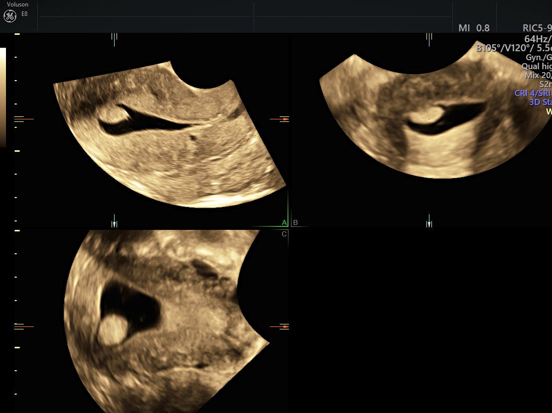
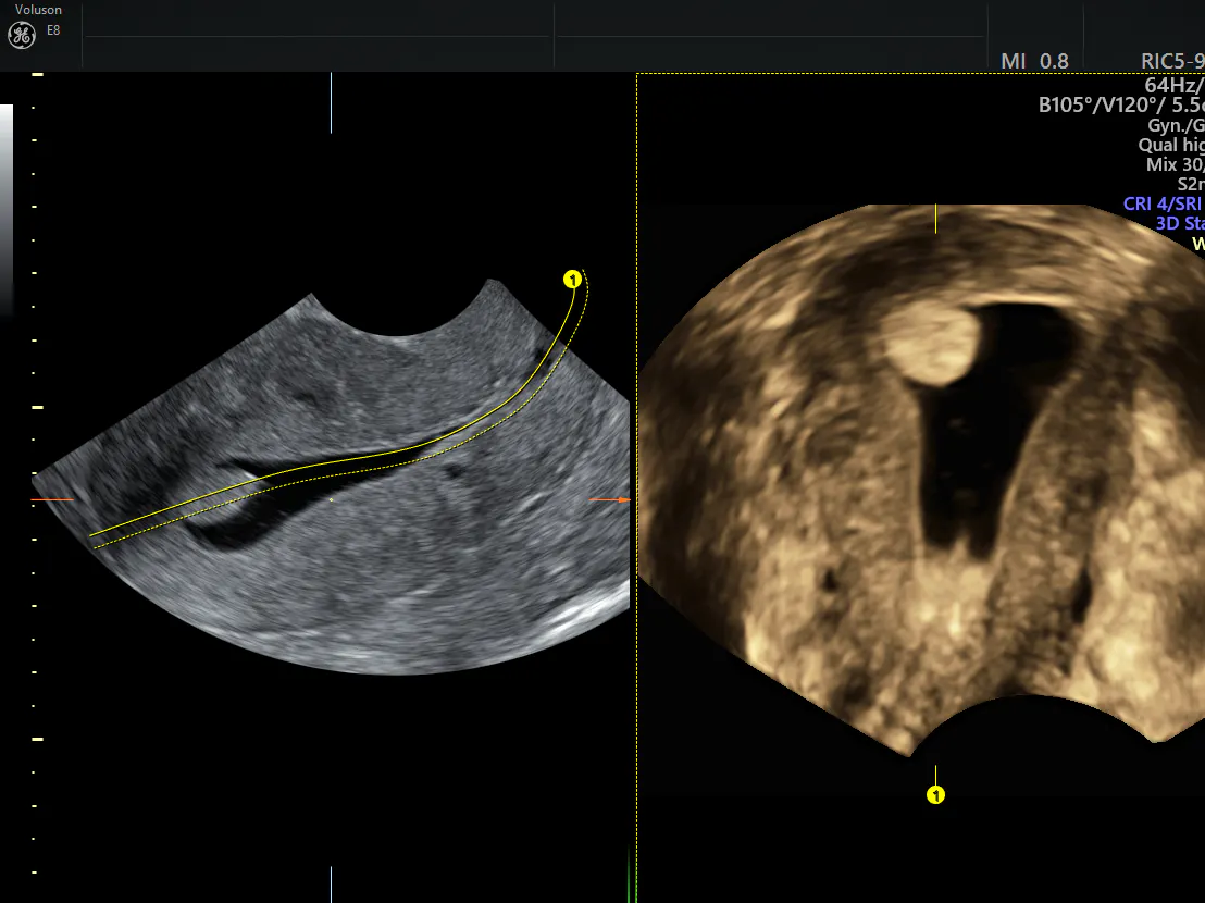
SIS with 3D endometrial polyp ultrasound is as simple and relatively painless as it was with 2D ultrasound. Sterile fluid is delivered to the uterine cavity via a slender catheter threaded through the cervix. The hypoechoic fluid outlines the polyp, which appears isoechoic to the surrounding endometrium. However, less fluid is needed to distend the cavity when combined with 3D ultrasound rather than 2D, shortening the duration of the procedure and improving acceptance by the patient.
endometrial polyp ultrasound rendering
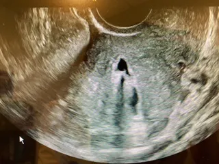
Image courtesy Dr. Lisbet Hanson
3D endometrial polyp ultrasound image
Collaboration Drives Better Decision-Making
The high-resolution images and cine loops can be stored or shared via Tricefy, a web-based image routing system that allows unlimited files to be archived and managed securely. It is HIPAA-compliant and facilitates collaboration with colleagues, providing images of superior definition for review, rather than dark or blurry printed photos. These images can even be shared with patients on any mobile phone, laptop or computer, thus meeting present-day expectations for rapid and decisive healthcare delivery.
Technology is revolutionizing clinical workflow, how physicians interact with their colleagues and patients, and what patients expect. Adaptation to change can be difficult and sometimes triggers myriad responses, but transitioning from 2D to 3D with SIS is an easy reach, not a quantum leap.

