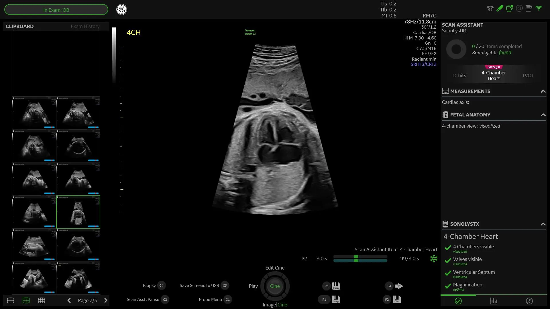
The Role AI Models Can Play for Ultrasound
Artificial intelligence (AI) is emerging as a key tool for ultrasound, particularly in prenatal diagnosis of congenital heart defects (CHDs). AI models can assist in raising the detection rate of congenital heart defects via prenatal ultrasound and have the potential to improve outcomes for babies born with CHDs.
CHDs are the most common of all birth defects, according to the Centers for Disease Control and Prevention. About one in four babies born with a heart defect has a critical CHD, meaning they need surgery or other procedures in the first year of their lives, says the CDC.
Training AI Models to Identify Ultrasound Images
Developing AI tools for ultrasound requires first training the AI system to correctly identify the images it will see. This is accomplished by feeding the algorithm immense amounts of data so that it can learn to recognize patterns such as the anatomy of an organ.
Aris Papageorghiou, research director of the Oxford Maternal and Perinatal Health Institute, explains that big data is critical to training neural networks. "You feed the computer lots of labeled images and over time it learns to recognize certain features [of those images]," he says. "It learns in a very similar way to the way we [humans] learn," he says. "It uses trial and error, moving through different layers of convoluted neural networks to find the optimal solution."
Machine learning can be applied to medical imaging as well, says Papageorghiou. Deep learning has been shown so far to be particularly effective at recognizing ultrasound image patterns—often as good as humans. Applying AI to ultrasonography is not difficult because there is an abundance of data: 30% of the world's data being generated is healthcare related. "It's estimated we use only about 4% to 5% of that data," he says. Training AI systems to identify medical images is possible on that front.
The AI systems are trained on an initial set of data which is then closed and "learning mode" is ended. Regulatory authorities in multiple countries prohibit machine learning systems from learning continuously on the new data they are exposed to during the course of their use. Systems may be updated occasionally to new data sets, but that will be a separate process. Papageorghiou notes that it's critical that AI developers have a strong clinical understanding of what the system should do. Technical capability is also important, but knowing what is important to sonographers and what will help improve their experience the most is the key.
Current Detection of Fetal Abnormalities via Ultrasound
In the setting of prenatal healthcare, better screening is the goal of ultrasonography. When using ultrasound for screening for fetal abnormalities, rates of detection vary. For some abnormalities, the rate is high and ultrasound picks up the vast majority of them. For fetal CHDs, the detection rate is as low as 45%. In most of the cases where abnormalities were missed, the heart scan was inadequate and/or incomplete. In only 50% was there effective use of magnification. These types of errors show where AI can potentially be used to improve the detection rate of CHDs.
The Use of AI to Improve Detection of CHDs
The system that Papageorghiou developed includes an AI "assistant" that is embedded in the software, reminding the ultrasonographer of anything they might otherwise miss. A quality score is assigned to each image. There is a list of structures that must be ticked off to ensure a complete and adequate scan. The AI assistant recognizes structures in an automated way and integrates measurements into the interface. The ultrasonographer can manipulate the calipers to take a manual measurement if the measurement seems off, but this tool can greatly increase the speed of a scan.
AI ultrasound systems can support quality control during screenings by ensuring that each scan is high quality and complete. The quality metrics are supported in every single view, and they are based on protocols recognized by the International Society of Ultrasound in Obstetrics & Gynecology (ISUOG) and the American Institute of Ultrasound in Medicine (AIUM). The AI system was designed to work automatically in the background so that ultrasonographers don't have to do anything to invoke its abilities. It is integrated into the system's user interface seamlessly, providing both quality metrics and measurements that can be adjusted manually as needed but are produced with minimal interaction.
In this way, "AI systems can support quality control during screening, acting as a peer reviewer on your shoulder and helping ensure that your scan is complete and adequate," says Papageorghiou. The ultrasonographer can still annotate the images as they would ordinarily, but the AI system is ensuring the completeness and thoroughness of the ultrasound exam.
In areas where there are too few ultrasonographers or where healthcare systems are reliant on transient populations of ultrasonographers who may have been trained differently, an AI system that assists in scans can help ensure that the quality within the department remains high and that they are able to meet the demand for scan appointments.
The Future of AI Models in Ultrasonography
AI and deep machine learning can be applied to many areas of ultrasonography, with improvement of fetal scans for CHDs just one possible application. Radiomics is another application that involves identifying differences that may predict health issues, such as a particular type of cervical image that is associated with preterm birth. Other AI models are being developed to estimate lung maturity and gestational age, and those are becoming reasonably good, says Papageorghiou.
AI models still suffer from false positives, which can be a real detriment in a prenatal screening setting. In the next several years, Papageorghiou says, reducing the rate of false positives will make the systems more useful. While AI will never replace trained sonographers or radiologists, it is poised to become a critical tool that will improve detection rates of congenital heart defects and other fetal abnormalities.
View Professor Aris Papageorghiou’s full lecture here: The Future of AI in fetal medicine.

