
The majority of cervical polyps are pretty unmistakable. These small, fleshy, cherry red protrusions of the endocervix are often found incidentally during a gynecology exam or ultrasound. A masterful twist of the ring forceps, and the cervical polyp is deftly removed.
Still, upon opening a speculum, most gynecologists have at least occasionally wondered, "What is this?" The growth may be unusually large; its origin and blood supply may be in question. It may even have some alarming features potentially indicative of a malignancy. But don't panic — chillax and proceed with caution. With a 3D ultrasound system, you can complete a prudent evaluation right from your office.
What Are Cervical Polyps?
Whether broad-based or thin-stalked, cervical polyps can originate from the surface or the canal of the cervix. They can occur at any age, but are more commonly present in women in their fourth and fifth decades of life. Smaller polyps of the cervix are typically 1 to 2 centimeters in size, while very large ones may fill or spill out of the vagina.
There is significantly more concern for an associated malignancy of the endometrium when found in postmenopausal symptomatic women. On rare occasions, what appears to be a simple cervical polyp may be a malignancy masquerading as a polyp. Adenosarcomas, tubulosquamous polyps, sarcoma botryoides and even non-Hodgkins lymphoma of the cervix can present as an innocent polyp of the cervix. Fortunately, these represent the exception rather than the rule. More commonly, what may appear to be a cervical polyp is actually a prolapsing endometrial polyp or fibroid.
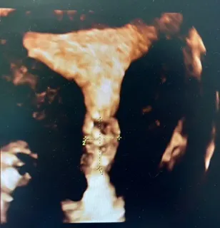
Polyps appear on ultrasound as elongated structures. A color Doppler image shows a single feeding vessel that typically runs the length of the polyp. They can look complex, with one or several cysts, or they can appear quite homogeneous. Fibroids, on the other hand, tend to be spherical and have a blood supply that runs near and around the surface of the structure. Fibroids appear similar in echogenicity to myometrial tissue.
Evaluating Cervical Polyps With Ultrasound
In every situation, it is critical to identify the origin of the feeding vessel in the pedicle. Beware: If the pedicle is very thick or vascular and appears to originate high in the endocervix or uterus, a twist of the ring forceps may not be the best solution. This is where transvaginal ultrasound (TVUS), 3D and 4D imaging of the cervix and uterus, saline contrast sonohysterography (SCSH) or sonovaginography can help sort this puzzle out.
The patient may be entirely asymptomatic. Typical complaints include abnormal and postmenopausal vaginal bleeding, menorrhagia, post-coital bleeding or vaginal discharge. If there is bleeding at the time of the TVUS, there may be no need for saline contrast because the blood acts as the contrast. Simply put on color Doppler and identify the feeding vessel.
SCSH is otherwise a safe and well-tolerated imaging tool that will allow evaluation of the endocervical canal as well as the endometrium. The only absolute contraindications are very heavy bleeding and pregnancy. With SCSH, a thin catheter is threaded through the endocervix and a small balloon is inflated to keep the catheter from being dislodged. Fluid is gently injected to distend the endometrial cavity while a TVUS is performed. To image the endocervical canal, the catheter can either be gently advanced, allowing fluid to flow into the endocervix, or the balloon can be let down and the fluid from the cavity passively drained while a cervical polyp ultrasound is performed. 3D ultrasound rendering with OmniView allows easy scrolling through the uterus and cervix for a virtual hysteroscopy without an invasive surgical procedure.
Sonovaginography with ultrasound gel to distend the vagina, coupled with transvaginal, transperineal or abdominal ultrasound, may be particularly useful for identifying polyps of the cervix. Patients with a low tolerance for vaginal exams may find this method much more acceptable than TVUS or SCSH.
Choosing 3D Cervical Polyp Ultrasound
Choosing the Right Procedure
Once you have identified the origin and blood supply, counsel your patient and schedule the appropriate procedure based on the information gathered. You may be able to proceed confidently with a quick office procedure, twisting off the pedicle with your Kelly or ring forceps until the polyp separates from its attachment. Apply some Monsel's solution to the base to achieve adequate hemostasis.
Alternatively, you may come to realize that you need to schedule a more intricate procedure under anesthesia at a convenient time — and not urgently after a failed attempt to remove what appeared to be an innocuous cervical polyp. Either way, be sure to send the tissue to pathology for documentation, and your investigation will be complete and thorough.

