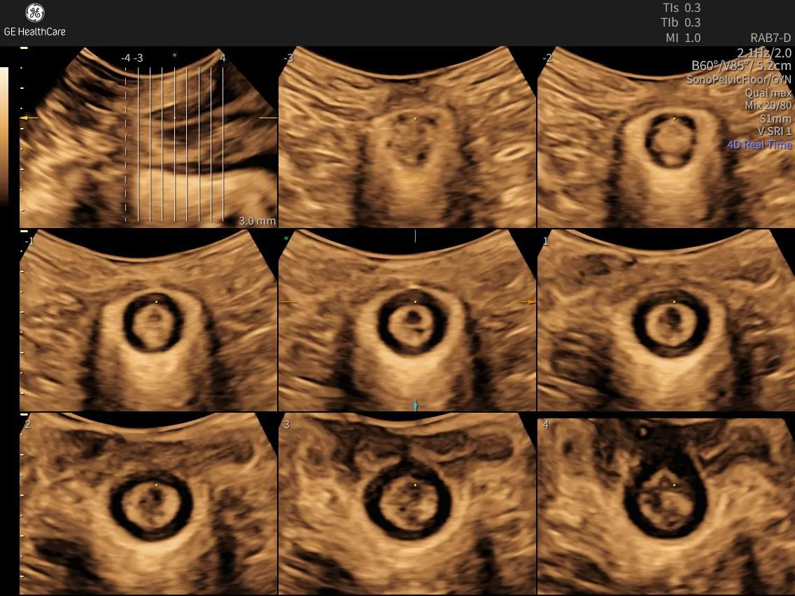
The Value of Adding a 360-Degree Probe to the Pelvic Floor Exam
Pelvic floor disorders affect millions of women worldwide, with conditions such as urinary incontinence, faecal incontinence, and pelvic organ prolapse impacting nearly one in four women during their lifetime.¹ Despite their prevalence, evaluating pelvic floor dysfunction remains challenging due to the complexity of the anatomy involved and the expertise required to accurately assess it.
A 360-degree endoanal/endovaginal probe, combined with a structured examination and reporting workflow such as Voluson™ SonoPelvicFloor, offers clinicians a more comprehensive approach to pelvic floor assessment. Importantly, the technology is only part of the equation. When paired with clinical expertise and a standardised imaging protocol, it can help clinicians build confidence, improve consistency, and strengthen diagnostic decision-making.
Understanding the Limitations of Standard Pelvic Floor Imaging
Transvaginal and transabdominal ultrasound are widely accepted as first-line imaging modalities for evaluating the female pelvis. However, assessing the pelvic floor presents unique challenges.
Structures such as the levator ani, puborectalis muscle, anal sphincter complex, and surrounding connective tissues form a highly interconnected three-dimensional system that may not be fully characterised using conventional imaging planes alone.
These limitations can have important clinical implications. During postpartum assessment, evaluation of incontinence, or preoperative planning for pelvic floor repair, standard imaging may not always capture the full extent of sphincter injury or levator trauma.²
Accurate characterisation matters. Misidentifying the severity of a sphincter defect or levator avulsion may influence treatment decisions, potentially affecting whether a patient is considered for conservative management or surgical intervention.
Comprehensive visualisation of the entire anal sphincter complex can provide valuable information when evaluating postpartum injury, assessing structural contributors to pelvic floor dysfunction, or determining the extent of fascial defects.
A Circumferential View of Complex Anatomy
The BK Medical/GE HealthCare 360-degree endoanal/endovaginal probe is designed to address many of the imaging challenges associated with pelvic floor assessment.
Using a rotating transducer, the probe generates a complete circumferential cross-sectional view of the anal canal and surrounding pelvic floor structures in a single acquisition. This enables clinicians to evaluate anatomy from every angle without the need for repeated probe repositioning.
The resulting circumferential perspective supports differentiation between internal and external anal sphincter defects, an important distinction when determining appropriate management pathways.
By capturing a continuous view of the anatomy, clinicians can localise defects according to both depth and position with a level of precision that may be difficult to achieve consistently with traditional 2D imaging alone.
The result is not simply enhanced visualisation, but the opportunity for more informed clinical assessment and decision-making.
Building Diagnostic Confidence with Voluson™ SonoPelvicFloor
High-quality pelvic floor imaging requires more than advanced hardware. Consistent image acquisition, accurate anatomical identification, and standardised reporting are essential components of a reproducible examination.
Voluson™ SonoPelvicFloor provides a protocol-driven workflow designed to guide clinicians through a structured pelvic floor assessment. By supporting a consistent examination methodology, the application helps reduce variability between operators and examinations while reinforcing pattern-recognition skills that are fundamental to interpreting pelvic floor pathology.
This structured approach is aligned with published consensus recommendations for pelvic floor ultrasound standardisation, including guidance from the International Urogynecological Association and International Continence Society (IUGA/ICS) and the American Institute of Ultrasound in Medicine (AIUM). These organisations emphasise the importance of reproducible acquisition protocols and structured reporting within clinical pelvic floor imaging.³
Clinical Applications for 360-Degree Pelvic Floor Imaging
The addition of a 360-degree probe expands the range of clinical questions that can be addressed during office-based pelvic floor evaluation.
Key applications include:
- Postpartum sphincter injury assessment
- Evaluation of faecal or urinary incontinence
- Assessment of pelvic organ prolapse
- Preoperative mapping for pelvic floor repair procedures
- Post-sphincteroplasty follow-up⁴
Levator ani muscle avulsion is identified on transperineal and endovaginal ultrasound in approximately 11% to 18% of women undergoing evaluation for pelvic floor dysfunction.⁵
The ability to identify structural trauma at the point of care can support more efficient clinical pathways, allowing clinicians to move directly from assessment to management planning without requiring additional imaging studies such as magnetic resonance imaging (MRI).
The Importance of Education and Clinical Expertise
Technology alone does not create diagnostic confidence.
A 360-degree probe may produce detailed images, but meaningful clinical insights depend on the expertise of the clinician acquiring and interpreting those images.
Developing confidence in pelvic floor ultrasound requires structured education, practical experience, and ongoing clinical development.
This is where GE HealthCare's commitment to clinician education plays an important role.
Through the Voluson Club, clinicians have access to online educational resources and hands-on learning opportunities designed to support continued professional development. Training opportunities include:
- Case-based learning modules
- Scanning technique tutorials
- Application-specific training for Voluson™ SonoPelvicFloor
- Guided learning pathways for pelvic floor ultrasound interpretation
These resources are designed to support obstetrician-gynaecologists, urogynecologists, colorectal surgeons, and ultrasound professionals as they progress from foundational anatomical understanding to advanced pattern recognition and clinical interpretation.
When education is combined with a structured workflow and advanced imaging technology, clinicians are better positioned to develop the expertise needed for confident and consistent pelvic floor assessment.
Connecting Advanced Imaging with Clinical Competency
The value of incorporating a BK Medical/GE HealthCare 360-degree endoanal/endovaginal probe into the pelvic floor examination extends beyond the technology itself. Its true value lies in enabling clinicians to visualise anatomy more comprehensively and evaluate pathology with greater confidence.
When paired with the structured workflow provided by Voluson™ SonoPelvicFloor, circumferential imaging can help address diagnostic gaps that traditional 2D imaging may not fully capture.
As with all advanced ultrasound applications, achieving the greatest benefit depends on the clinician's knowledge of anatomy, mastery of scanning techniques, and ability to integrate findings into patient management.
That combination of technology, education, and expertise helps transform imaging into meaningful clinical insight, supporting confident decision-making and advancing the standard of pelvic floor care.
References:
1.U.S. Food and Drug Administration Office of Women's Health. May 2025 - Know Your Pelvic Floor. FDA. May 2025. Accessed July 10, 2026. https://www.fda.gov/consumers/knowledge-and-news-women-owh-blog/what-women-need-know-about-their-pelvic-floor
2.Fritel X, Pizzoferrato AC, Letouzey V, Legendre G, de Tayrac R, Jundt K, Fauconnier A. The role of imaging in assessing perineal trauma. In: Perineal Trauma at Childbirth. Springer; 2016:115-145. doi:10.1007/978-3-319-14860-1_7
3.AIUM/IUGA Practice Parameter for the Performance of Urogynecological Ultrasound Examinations: Developed in Collaboration with the ACR, the AUGS, the AUA, and the SRU. J Ultrasound Med. 2019;38(4):851–864. doi:10.1002/jum.14953
4.Santoro GA, Colangelo AC, Pelizzo P, Cian R, Zanus G. Imaging modalities for pelvic floor disorders. Ann Laparoscopic Endosc Surg. 2022;7:13. doi:10.21037/ales-21-44
5.van Gruting IMA, Stankiewicz A, van Delft KWM, Doumouchtsis SK, Inthout J, Sultan AH, Thakar R. Diagnostic test accuracy of magnetic resonance imaging and pelvic floor ultrasound for diagnosis of levator ani muscle avulsion. Ultrasound Obstet Gynecol. 2022;60(4):559–569. doi:10.1002/uog.24955

