Ultrasound Scanning: Why and How to Move From 2D to 3D Ultrasound
Ultrasound scanning is invaluable for diagnosing pathologies that may prevent IVF success. Dr. Nick Raine-Fenning, an expert in IVF at the University of Nottingham, shares his experiences using 2D and 3D ultrasound with IVF patients and explains why he considers 3D a staple of his practice.
Dr. Raine-Fenning begins his lecture by explaining his approach to each new IVF patient, which involves the following questions:
- Is there regular and predictable ovulation?
- What's in the patient's ovarian reserve, and what factors might influence egg collection?
- Are there any pathologies of the fallopian tubes?
- Is the endometrium favorable for implantation?
Assess Ovulation
While 3D ultrasound scanning is not strictly necessary for every exam, Dr. Raine-Fenning's practice always uses 3D, in order to have the most comprehensive information available for the file. In determining the best imaging solution for ovulation assessment (Question 1), he states, "3D doesn't really add much. We can see a dominant follicle develop, and then that will disappear and be replaced by the corpus luteum...which is [easily visible in greyscale]." In fact, his practice doesn't use ultrasound at all to assess ovulation. Instead, they use progesterone measurements in patients with regular cycles and assume that patients with irregular cycles don't ovulate.
Evaluate Ovarian Reserve
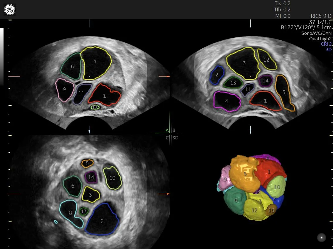
While clinicians don't need ultrasound to evaluate ovulation, the tool is critical when assessing ovarian reserve. Though some debate the value of using the anti-Mullerian (AMH) marker instead of ultrasound for antral follicle count, Dr. Raine-Fenning prefers a scan for antral follicles in this case. Clinicians sometimes dismiss antral follicle count as a test for ovarian reserve, he explains, as the slightest user error (the wrong angle, frame rate, frequency, or harmonics) may diminish image quality. Making the proper adjustments ensures the follicles stand out, even with reduced image clarity.
Dr. Raine-Fenning makes several salient points about the power of ultrasound machines and the need for user proficiency. "When you're looking for the follicle, focus on the follicle," he says. "Then, when you're looking at the uterus, focus on the uterus." The stenographer, he stresses, must use different settings, from the left to the right ovary, and then to the uterus and the follicles.
To capture a useful image, the sonographer needs the hypoechoic fluid to stand out in the follicles. They can accomplish this by paying attention to frame rate, angle, definition, zoom, resolution, and gain. Again, it's important to adjust the settings for each ovary.
Personalize Ovarian Stimulation
Dr. Raine-Fenning discusses patients undergoing ovarian stimulation; the amount of stimulation depends on the number of follicles available. Patients with a low number of follicles on ultrasound examination receive a high dose of stimulation in order to retrieve a higher number of eggs. A patient with a high number of follicles (e.g., experiencing polycystic disease) would be approached with care and administered a lower dose of stimulation. Meanwhile, a patient with an average number of follicles would get an average dose, with a goal of retrieving around 15 eggs. For all patients, using ultrasound appropriately is essential to provide quality images and ensure that follicles are not over- or under-counted.
Clinical Value of 3D Ultrasound
Dr. Raine-Fenning reflects, "I'm often asked, and it's a really important question, is 3D still 20 years down the line?" Practitioners wonder whether 3D is a "gimmick" or if it adds value in clinical practice. His answer is consistent: obtaining a 3D volume allows you to interact with that image or data "in a way that you cannot interact with a static image."
As an example of 3D utility in clinical practice, Dr. Raine-Fenning mentions measuring the volume of follicles based on diameter. Though, as non-spherical objects, follicles technically lack diameter, the SonoAVC™ tool "imagines" the follicle as a sphere to measure its diameter and obtain its volume.
3D ultrasound is also valuable as a teaching tool and in a research setting.
Detect and Diagnose Pathologies
Turning to pathology, Dr. Raine-Fenning displays a scan of a hydrosalpinx, which he refers as the nemesis of fertility physicians. While the featured image clearly reveals the hydrosalpinx, this isn't always the case. However, clinicians need to understand this important pathology before proceeding with the patient's treatment. If it is indeed a hydrosalpinx, it needs to be removed. Volume is great, but for adnexal pathology, he prefers tomographic ultrasound imaging, because it is easy to apply.
For uterine pathology, Dr. Raine-Fenning demonstrates how to distinguish uterine polyps, which frequently delay IVF treatment. He suggests obtaining a volume scan, which can be scrolled through in detail to diagnose the type of polyp and plan the surgery. He adds that a saline infusion, sometimes necessary during a volume scan, is often made obsolete by use of 3D.
Although physicians are often more worried about the presence of submucosal fibroids than mural fibroids, the presence of a mural fibroid of 2cm or more, whether or not it has cavity involvement, can negatively affect the IVF process. This doesn't always lead to surgery, Dr. Raine-Fenning says, but it does underscore the importance of the diagnosis in determining whether an intervention is needed.
Using volume ultrasound when evaluating endometrial pathology can enable the physician to perform a virtual hysteroscopy with the 3D rendering tool. They can also detect adenomyosis by observing changes to the endometrial/myometrial border, as the border itself becomes unclear. Other features better seen with 3D include increased echogenicity, hypovascularity, and subendometrial cysts.
Dr. Raine-Fenning suggests using OmniView to accurately diagnose uterine anomalies such as bicornuate, unicornuate, and didelphic uteri. He describes these anomalies as "nonsurgical cases" and stresses the importance of informing patients of the high-risk nature of their pregnancies. He also shares surprising results from a recent study, which found that surgical resection of a uterine septum did not increase conception rates or live births.
Benefits of 3D Ultrasound Scanning in IVF Practice
Practitioners may wonder, "Why 3D?" Dr. Raine-Fenning's response is, "Why not?" He says most sonographers are likely already using a machine with 3D capability. While a 3D system may not be necessary with every single exam or view, it allows clinicians to capture significantly more data; measure it objectively, reliably, reproducibly, and with validity; and reduce inter-operator variability. Finally, he adds that physicians who come to his unit for ultrasound training do not begin with 2D scans; they start their education with 3D, which enhances their knowledge of pelvic anatomy and pathology as they learn how the machines work.
View Dr. Raine-Fenning’s full video ‘Taking your fertility assessment from 2D to 3D’.

