

Pelvic inflammatory disease (PID) will affect more than 1 million people in the United States this year, according to the American College of Obstetricians and Gynecologists. It can present in either acute or chronic forms, and it can be asymptomatic or include symptoms such as abdominal and pelvic pain, vaginal discharge, fever, painful intercourse or painful urination.
Without a timely and accurate pelvic inflammatory disease diagnosis, complications such as tubo-ovarian abscesses and fallopian tube scarring can lead to infertility, chronic pelvic pain and ectopic pregnancies. PID can be a silent disease with significant long-term consequences, so having a fast and effective tool to assess and diagnose PID is extremely valuable for clinicians.
The ideal diagnostic imaging exam performed in cases of suspected PID should be quick, thorough, readily available in doctors' offices and easily able to assess the pelvis, especially the ovaries and fallopian tubes. Ultrasound has been recommended as an effective modality for accurate and timely pelvic inflammatory disease diagnosis by a 2017 Medscape review.
What Happens During a PID Infection
A PID infection is caused by pathogens ascending from the cervix or vagina and spreading into the endometrium, fallopian tubes, ovaries and adjacent pelvic structures. The causes of PID can be inadequately treated chlamydia or gonorrhea, untreated STIs of any kind or douching shortly after an IUD is inserted.
The early stages of an infection bring cervicitis, oophoritis, salpingitis, thickening of the uterosacral ligaments and accumulation of simple fluid in the endometrial canal, fallopian tubes (hydrosalpinx) and pelvis. As the disease progresses, the fallopian tubes become thicker and fill with complex fluid, becoming a pyosalpinx. Later, tubo-ovarian and pelvic abscesses form thick-walled, complex fluid collections that may contain internal septations, a fluid-debris level or, less commonly, gas. The presence of gas is a definite sign of infection.
The tubal scarring caused by PID can result in an infertility diagnosis, chronic pelvic pain and ectopic pregnancy, and complicated PID may even contribute to patient mortality.
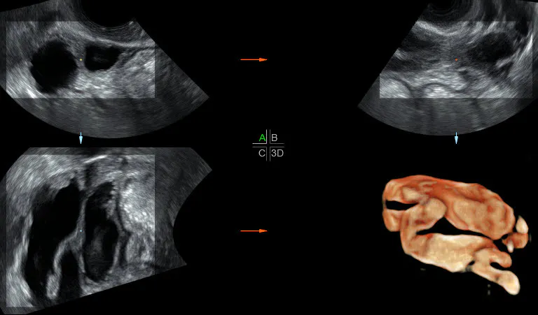
Going 3D: Pelvic Inflammatory Disease Ultrasound Exams
In acute PID, the ovaries can be enlarged with an increased number of follicles, and the fallopian tubes can dilate with thickened walls, increased vascularity and formation of a complex, ill-defined, multi-locuated structure known as a tubo-ovarian abscess (TOA). An untreated TOA can rupture, resulting in peritonitis and further infection spread.
In chronic PID, a hydrosalpinx can form from a fluid accumulation within a scarred and blocked fallopian tube, and a peritoneal inclusion cyst may develop around the uterus and ovaries, appearing as a loculated fluid collection.
PID Ultrasound Hydrosalpinx
These complex, fluid-filled structures are well-suited for evaluation by PID ultrasound exams. Given that 3D ultrasound uses a wide-field view, digitally reconstructed slices and a third scanning plane (the coronal plane), it can provide a more detailed view of the pelvis, uterus, fallopian tubes, ovaries and both adnexa. This extra information allows for a more accurate pelvic inflammatory disease diagnosis and can help to prevent women from suffering from the long-term effects of PID.
PID Ultrasound Images: Making Diagnosis Easier
A connected ultrasound system is another very helpful feature for the PID patient. Using systems such as GE's Voluson with embedded Trifecy tool is helpful for sharing images with colleagues to develop treatment plans and for getting a second opinion on a complex collection of pelvic pathology.
Don't be stumped again by complex acute or chronic PID. With 3D ultrasound, there's a better way to assess and diagnose.

