The history of ultrasound in obstetrics and gynecology dates back to a foundational paper published in The Lancet in 1958 by Ian Donald of Glasgow, Scotland. Technological developments since then have led to much more advanced forms of ultrasound available today, including real-time imaging, transvaginal probes, color, and power Doppler and 3D and 4D imaging. Although gynecologic ultrasound exams originally took place later in pregnancy, first-trimester ultrasound is now widely used for a number of diagnostic and therapeutic indications.
How First-Trimester Ultrasound Came About
Once real-time transvaginal ultrasound became available in the mid-1980s, researchers rapidly began to find new applications for it. Dr. Ilan Timor-Tritch from Columbia University pioneered the concept of transvaginal sonoembryology and used ultrasound to diagnose early embryonic abnormalities. Several researchers in the late 1980s also developed diagnostic criteria for ectopic pregnancy, leading to successful identification in more than 90 percent of cases.
Until the 1990s, the use of ultrasound in obstetrics was limited to the second and third trimesters. Measuring fetal nuchal translucency thickness during 11 to 13 weeks of gestation became part of a screening for Down syndrome (trisomy 21) thanks to the research of Dr. Beryl Benacerraf in Boston and Prof. Kypros Nicolaides from King's College, London. This development ushered in the regular use of first-trimester ultrasound. Nicolaides has since established a certification program for nuchal translucency.
In 1986, Prof. Benacerraf and her group in Boston first described an increased skin fold measurement in the second trimester associated with Down syndrome. In 1992, Dr. Nicolaides learned that the excess skin found in individuals with Down syndrome could be visualized on ultrasound as early as the third month of fetal development via increased nuchal translucency.
When considered alongside maternal age and maternal serum free ß-human chorionic gonadotropin and pregnancy-associated plasma protein-A measured during the same period, nuchal translucency thickness measurements can help detect certain chromosomal defects at a rate of about 85 to 90 percent. In 2001, researchers discovered that adding ultrasound examination of the nasal bone raises the detection rate to more than 95 percent.
Doppler and Preventing Pregnancy Risks
The advent of color Doppler as an integral part of ultrasound allowed for better evaluation of fetal circulation and cardiac function. Today, Doppler is still used to evaluate fetal circulation to assess fetal well-being and timing of delivery in the case of a compromised fetus.
Now, Prof. Nicolaides and others are exploring the possibilities of using ultrasound to screen for preeclampsia in the first trimester, using uterine artery Doppler in conjunction with biomarkers. Preventive therapy appears to be effective only if initiated during the first trimester, making early detection critical. Risk assessment for preeclampsia can be completed with the Fetal Medicine Foundation's preeclampsia risk calculator.
First-trimester ultrasound is also used for a number of medical indications unique to the earliest stages of pregnancy. Ultrasound is crucial for needle positioning in amniocentesis. It is also used to guide the needle to remove a cord blood sample to assess various biomarkers related to fetal health.
Early Fetal Ultrasound: Looking Ahead
Recent advances in ultrasound technology offer better resolution for finer details. The development of 3D ultrasound brought clinicians the ability to detect superficial fetal structural defects and secondary palate defects, as well as to monitor fetal cardiac activity and visualize more complex fetal cardiac anatomical relationships, potentially identifying fetal cardiac anomalies. Ultrasound training and education are also catching up, but first-trimester obstetric exams remain among the hardest to perform. For patients, better ultrasound leads to early diagnosis of fetal abnormalities which can help them make more informed decisions about their pregnancy.
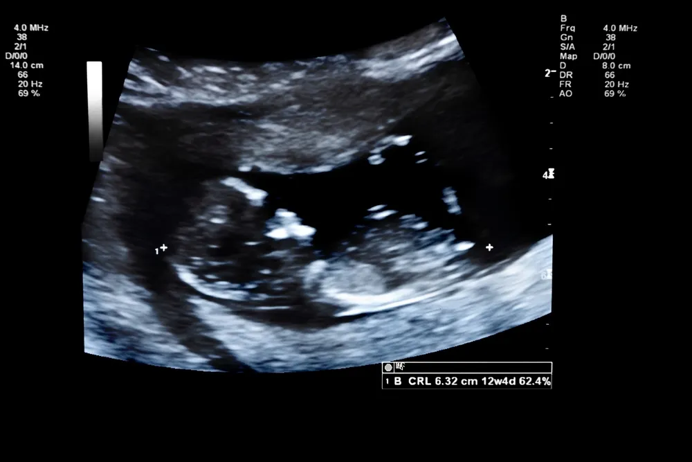
As we look at the future, the use of AI is increasing in prenatal ultrasound. In first trimester ultrasound, AI can help with detection and identificaton of fetal limbs, to assess fetal development, and to perform automatic measurement of nuchal translucency and crown-rump length.
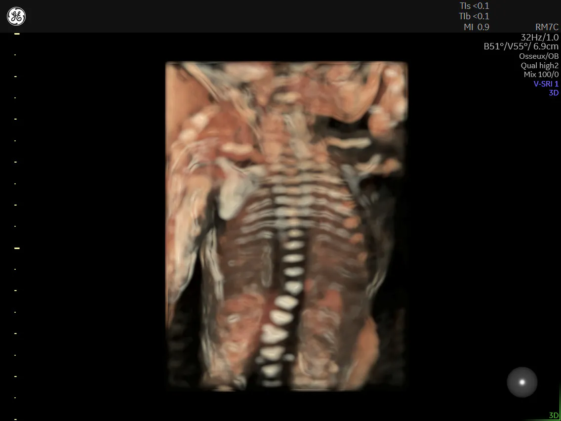
HDlive™ rendered coronal view of 18 week fetal spine.
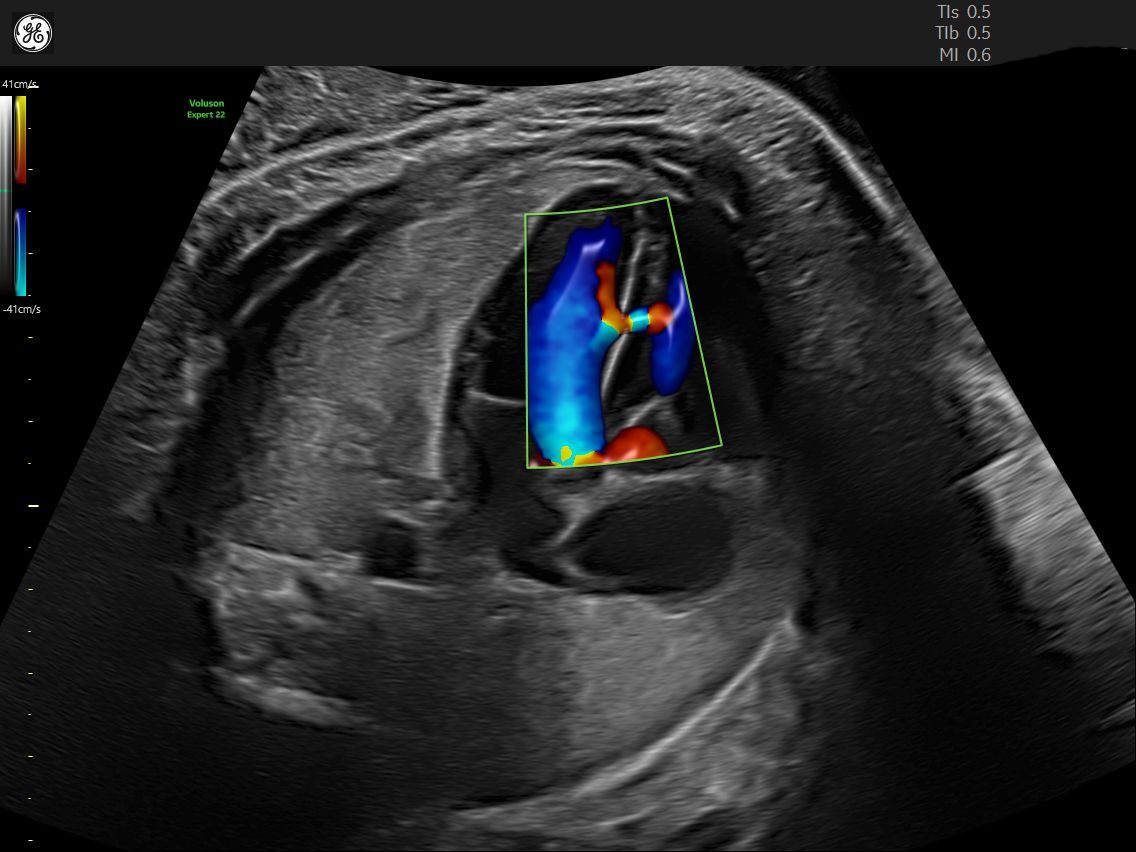
Radiantflow™ helps confirm a ventricular septal defect in 21 week fetal heart.
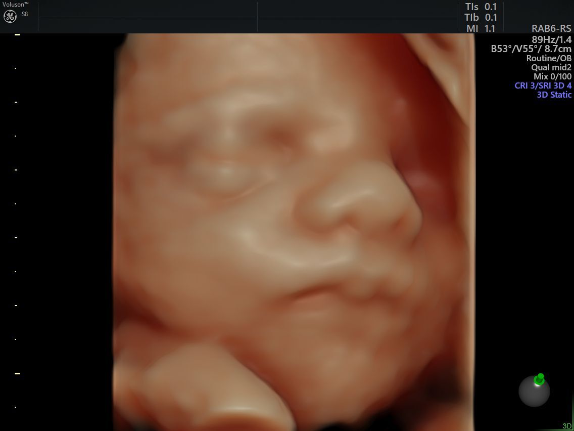
Fetal Face using Voluson HDlive™ technology
Ultrasound represents one of the biggest and most critical advances in obstetrics and gynecology in the 20th and 21st centuries. Its use has permeated all aspects of care, and OB/GYNs are still identifying new use cases. The history of ultrasound could—and does—fill a whole volume. Taking some time to review first-trimester ultrasound can provide helpful context as to how this technology was first developed, how it's used now, and how it can make a difference in the future.