
What is your personal best practice for checking intrauterine device (IUD) placement with ultrasound? Are you still relying on 2D ultrasound, or perhaps simply a pelvic exam, for a string check? If so, it's time to consider using 3D ultrasound to evaluate IUD placement.
3D sonography is an easy procedure that you can provide conveniently from your office. In this era of quality improvement and cost containment, this technology is relatively inexpensive, does not expose the patient to ionizing radiation, and is much less invasive and more accurate than other options.
How to Check IUD Placement: New Solutions to Old Challenges
In decades past, physicians located IUDs by placing a metal sound in the uterine cavity and producing X-rays of the abdomen. Depending on when you began your training, you may have used that technique or relied on computed tomography (CT) scans, an expensive procedure with comparatively high radiation exposure. CT is still the choice for tangled intra-abdominal IUDs involving bowels, whereas magnetic resonance imaging has not traditionally been used for IUD localization.
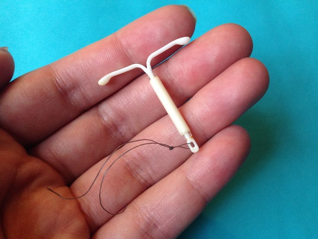
More recently, practitioners relied on 2D ultrasound to identify IUD placement. Unfortunately, given that IUDs are composed of different materials, 2D ultrasound does not always provide a complete visualization of the device. Some IUDs contain copper, which makes them partially radiopaque and hyperechoic on ultrasound. Others are made entirely of nonmetallic materials containing polyethylene, silicone and barium sulfate, which make them radiopaque but nonvisualizing on ultrasound. Additionally, embedded arms and twisted IUDs are difficult to identify with 2D ultrasound. Challenging anatomy can also impede verification.
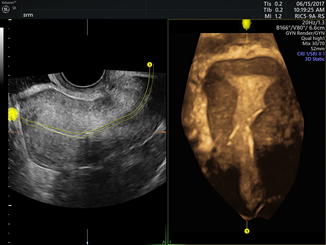
IUD placement ultrasound shows device is too low in the cavity with the right arm penetrating the myometrium
The 3D reconstruction, particularly the coronal view, sidesteps these problems. The images have been likened to a virtual reality experience that allows structures and their relationship to be compared to each other. This IUD placement ultrasound allows you to locate the device within the uterine cavity accurately, consistently and confidently.
Adopting 3D Ultrasound for IUD Placement
Learning to master new technology can be intimidating at first glance. Ultrasound is an innovative technology that has gained a global audience and enormous market share in the world of medical imaging. Today, it plays a vital role in gynecologic healthcare.
Training programs emphasize the skills required to operate a 3D ultrasound system. After residency, healthcare professionals must commit to a lifetime of self-learning to master those skills. Fortunately, ultrasound technology has become increasingly efficient and user-friendly as it has matured.
Change is always a gradual process. If you are upgrading from 2D to 3D ultrasound, the shift will be relatively seamless. Still, you should take basic steps to prepare yourself and your practice to adopt the new technology. Sign up for webinars or focused ultrasound courses with practical training in 3D. You might also consider viewing vendor demonstrations or working with a trusted mentor to brush up on the skills required to operate a 3D ultrasound system.
For a busy gynecology practice, 3D ultrasound is the best imaging technology for all IUD location evaluations. Rather than impede workflow, it improves efficiency and streamlines the patient experience.
If you have yet to consider adopting IUD placement ultrasound, now is a great time to start the process. A modest amount of effort can save your practice time and money, and it can help you forge a strong bond with your patients by improving their experience in your office.