

Between 30% and 50% of women with endometriosis are infertile.1 While the disease doesn’t cause infertility—it may make it more difficult for your patients to conceive.
Using Ultrasound to Connect Endometriosis and Infertility
Ultrasound Leads to Better Understanding in the Diagnosis and Management of Endometriosis Patients
Experts insist that ultrasound leads to more information and a better understanding of the diagnosis and management of endometriosis patients. Learn why.

Between 30% and 50% of women with endometriosis are infertile.1 While the disease doesn’t cause infertility—it may make it more difficult for your patients to conceive.
With endometriosis, it’s impossible to know your patient’s odds of getting pregnant until she tries. But this common condition is considered a negative influencing factor—and the likelihood of fertility issues increases with the severity of the disease. That’s why early diagnosis and management are critical for women who want to start a family, and experts insist that ultrasound leads to more information and better understanding.
“The most important thing is to diagnose the early disease, but it’s not easy because mild endometriosis is difficult to recognize. So, we have to be on alert if a patient has any symptoms—and learn to detect endometriosis from small signs that can be seen on ultrasound,” said expert gynecologist, Caterina Exacoustos, M.D. from the University of Rome.
Dr. Exacoustos favors high-quality ultrasound as a first-line modality in detection of endometriosis because she says laparoscopy can’t always be the gold standard for diagnosing the condition. She’s backed by a growing body of evidence that supports the use of ultrasound in the diagnosis and surgical planning for endometriosis.2 Studies show endometriomas can easily be detected through transvaginal ultrasound—and imaging can provide a comprehensive depiction of deep infiltrating endometriosis lesions in the pelvic and subperitoneal areas, not easily accessible laparoscopically.3
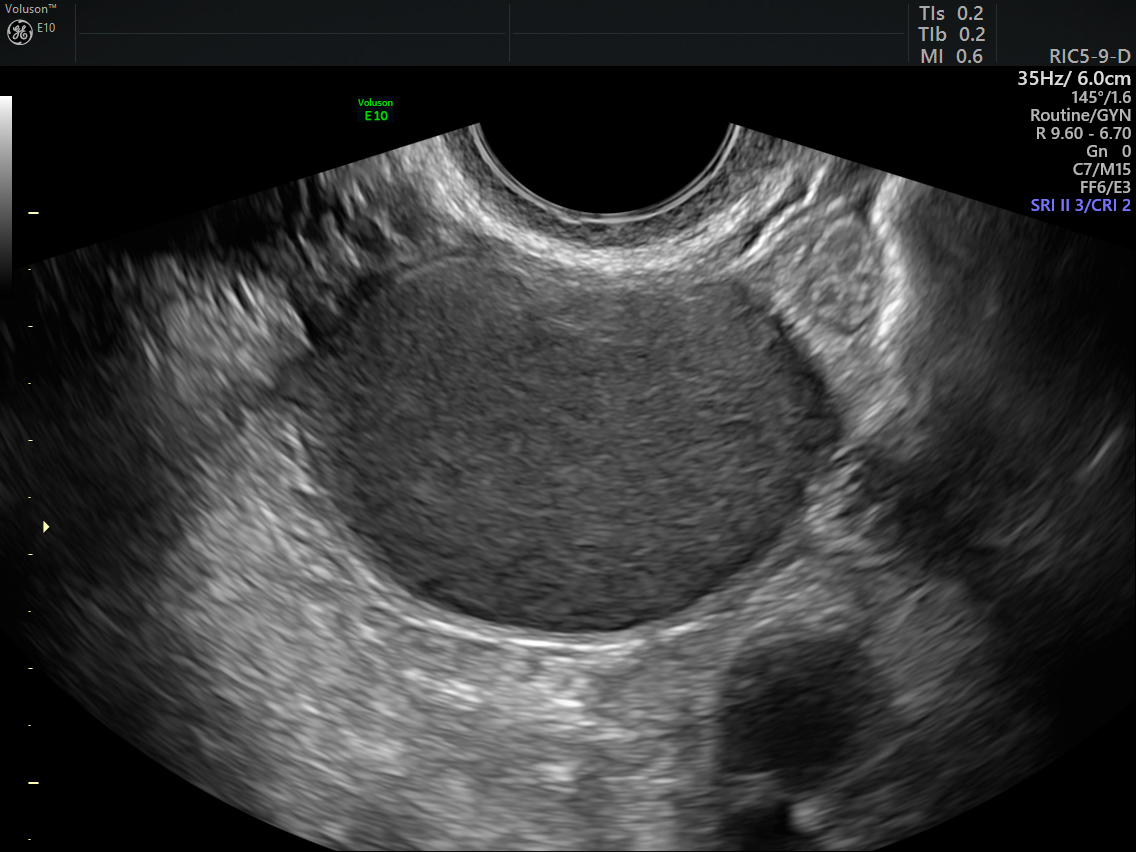
Transvaginal ultrasound image of endometrioma on ovary
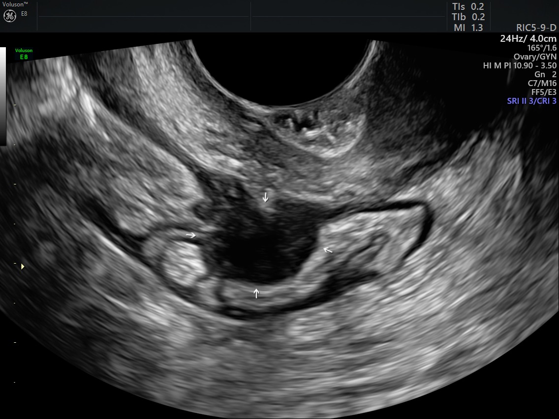
Deep Infiltrating Endometriosis (DIE) in bowel wall
“It’s the first tool that we can see inside the uterus, pelvis, bladder and rectum. You can see more with ultrasound than with laparoscopy, and it’s much more accurate. 3D is much better in the diagnosis of adenomyosis, which is a very important factor affecting fertility,” Dr. Exacoustos stressed.
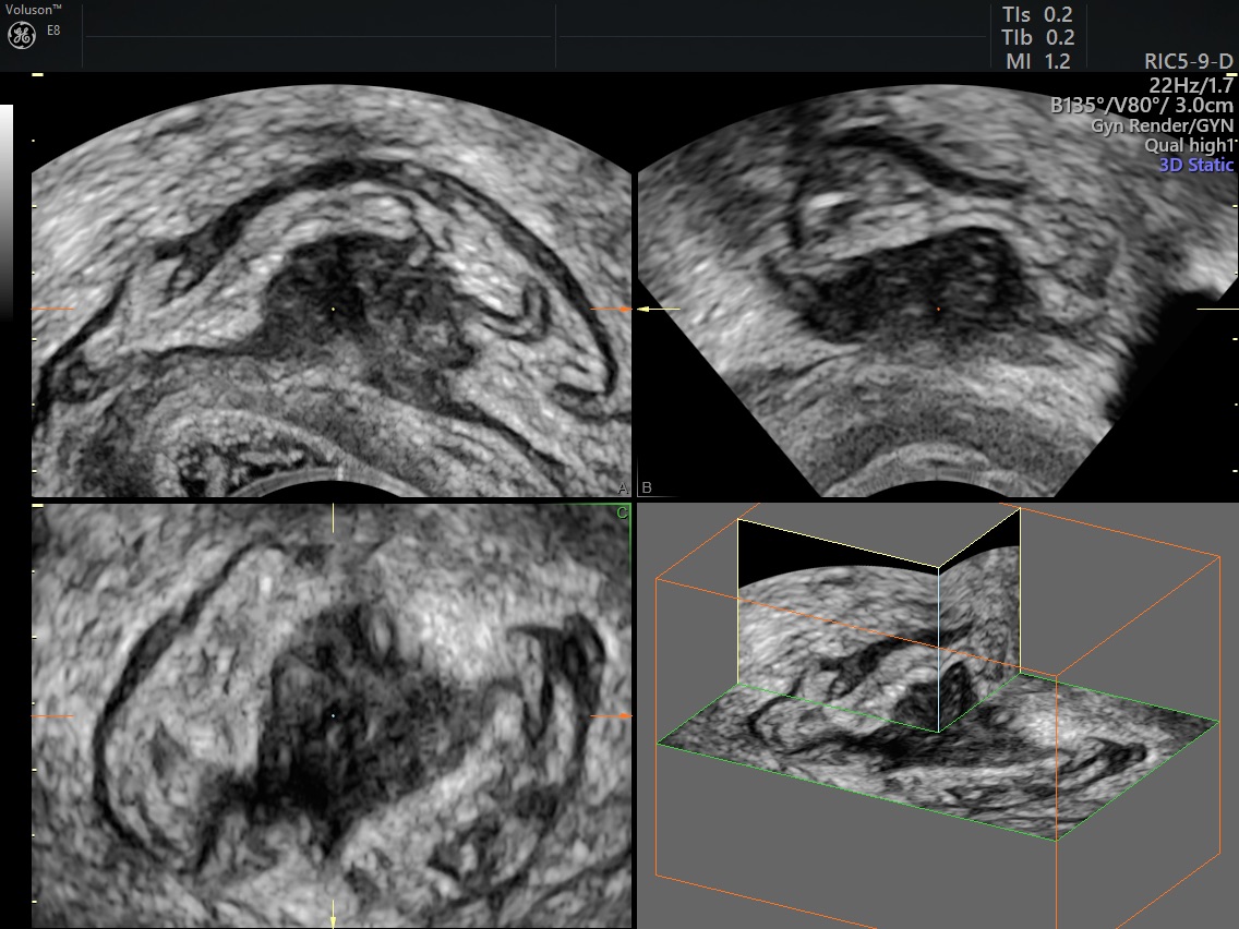
3D ultrasound volume of DIE
To help find answers, Dr. Exacoustos utilizes specialized tools available on Voluson™ technology. The ultrasound, by GE Healthcare, features Advanced VCI with OmniView—which can improve contrast resolution and visualization in any image plane, even irregularly shaped structures—making it easier to confirm a presumptive diagnosis. The Voluson systems also enable you to confidently evaluate the uterine cavity and determine tubal patency by performing detailed hysterosalpingo-contrast-sonography (HyCoSy).
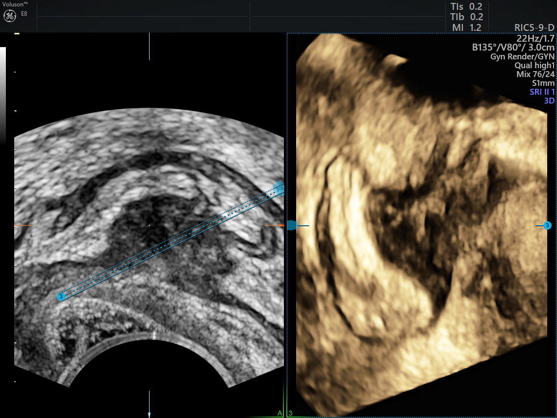
DIE seen with Advanced VCI with OmniView
When it comes to endometriosis, the power of ultrasound is also demonstrated in the real-time dynamic technique known as ‘sliding sign.’ By moving your probe, you can determine if the posterior cervix-uterus glides freely across the bowel. It indicates pouch of Douglas involvement and obliteration—which suggests DIE of the rectum. In fact, a systematic review and meta-analysis determined ultrasound was effective in the preoperative diagnosis of bowel endometriosis with pooled estimates of 91% sensitivity and 98% specificity.5
Dynamic movement confirms frozen endometrial implant in pelvis
That research was led by respected endometriosis and infertility surgeon, Gernot Hudelist M.D. P.D., M.Sc, of St. John of God Hospital in Vienna. He believes ultrasound offers valuable predictors that help ensure patients are treated in the right place, by surgeons with the right skills—for the best care.
“You have to triage patients with pelvic pain into extensive disease patients who have to go to tertiary centers versus patients with a minor disease who can be treated in other places. Transvaginal ultrasound allows a noninvasive picture that gives us a very quick and accurate idea of the possibility of extensive disease. It gives us a much better idea of the extent of the problem,” Dr. Hudelist said.
Ultrasound enables you to map out the disease and coordinate interdisciplinary procedures to optimize results. Imaging can also influence patient consent and help you predict possible outcomes.
“You will be able to tell the patient how high the chances will be to achieve a pregnancy if you do this or that. And if the surgery goes right—you can talk about what the main outcome will be regarding pain issues, what the outcome will be regarding spontaneous conception rates, what the outcome will be in case she will undergo IVF.”
Even with all the benefits, Dr. Hudelist says it’s rare for surgeons to perform ultrasound. He calls it ‘the missing link,’ and believes too many of his colleagues go into the operating room blind.
“I think there are a lot of surgeons who don’t know at the beginning what they’re going to get. Some of them are able and capable to solve it. Some of them are not. The approach should be to verify the possibility of disease non-surgically with ultrasound, to triage patients adequately, and to take them to theaters with a concept—with a plan.”
For Dr. Hudelist and Dr. Exacoustous, it’s simple. The more you know, the more you can prepare. And when a woman’s fertility is at stake—that information could change everything.
-
A Voluson 3D ultrasound of the pelvic anatomy, coupled with a careful exam, can help streamline the process of diagnosing endometriosis. Learn how Dr. Hanson manages her patients and sign up to Stay Informed.
-
Learn more about the 4-step ultrasound evaluation as suggested by the International Deep Endometriosis Analysis (IDEA) group.
-
When evaluating and diagnosing endometriosis, 3D ultrasound can deliver the visibility gynecologists need to create a personalized treatment plan
-
You can help your patients better understand the similarities and differences of endometriosis and endometrial cancer.
-
Endometriosis is notoriously hard to diagnose, but once it has been, there are various options for how to treat endometriosis which you can explore with your patient.
-
Orilissa is a recently approved endometriosis medication for treatment of moderate to severe endometriosis pain. Learn more about this endometriosis medication.
-
Are your patients asking about an endometriosis diet? Research shows anti-inflammatory foods and other nutritional choices may help alleviate pain.
-
Transvaginal ultrasound was found to be a reliable predictor of ureteral endometriosis in patients with known deep infiltrating endometriosis.
-
After a diagnosis, a woman will need to consult a number of endometriosis doctors, specialists and surgeons as part of treatment. Here’s what they can expect.
-
What is ultrasound used for? Ultrasound has many applications for women’s health. Read our collection of FAQs.
Macer ML, Taylor HS. Endometriosis and infertility: a review of the pathogenesis and treatment of endometriosis-associated infertility. Obstet Gynecol Clin North Am. 2012;39(4):535–549
Hoyos L, Benacerraf B, Puscheck E, Imaging in Endometriosis and Adenomyosis, Clincal Obestetrics and Gynecol. 2017 March;60(1):27-37
Kondo W, Zomer M.T., Pinto, E.P., Deep infiltrating endometriosis: Imaging features and laparoscopic correlation, Journal of Endometriosis. 2011;3. 192-212.
Reid S, LU C, Casikar I, Prediction of pouch of Douglas obliteration in women with suspected endometriosis using a real-time dynamic transvaginal ultrasound technique: the sliding sign, Ultrasound Obstet Gynecol. 2013 June;41(6):685-91
Hudelist G, English J, Thomas AE, Tinelli A, Singer CF, Keckstein J. Diagnostic accuracy of transvaginal ultrasound for non-invasive diagnosis of bowel endometriosis: systematic review and meta-analysis. Ultrasound Obstet Gynecol 2011; 37:257–263
Hudelist G., Oberwinkler K.H., Singer C.F., Tuttlies F., Rauter G., Ritter O. et al. Combination of transvaginal sonography and clinical examination for preoperative diagnosis of pelvic endometriosis. Hum Reprod 2009;24:1018-24;
Guerriero S, Condous G, van den Bosch T, Valentin L, Leone FP, Van Schoubroeck,D, Exacoustos C, Installé AJ, Martins WP, Abrao MS, et al . Systematic approach to sonographic evaluation of the pelvis in women with suspected endometriosis, including terms, definitions and measurements: a consensus opinion from the International Deep Endometriosis Analysis (IDEA) group. Ultrasound Obstet Gynecol. 2016;48:318-32
Exacoustos C, Malzoni M, Di Giovanni A, Lazzeri L, Tosti C, Petraglia F, Zupi E. Ultrasound mapping system for the surgical management of deep infiltrating endometriosis. Fertil Steril 2014;102:143-150.
Exacoustos C, Manganaro L, Zupi E. Imaging for the evaluation of endometriosis and adenomyosis. Best Pract Res Clin Obstet Gynaecol 2014;28:655–81
Exacoustos C, Manganaro L, Zupi E. Imaging for the evaluation of endometriosis and adenomyosis. Best Pract Res Clin Obstet Gynaecol 2014;28:655–81
Lazzeri L, Morosetti G, Centini G, Monti G, Zupi E, Piccione E, Exacoustos C. A sonographic classification of adenomyosis: interobserver reproducibility in the evaluation of type and degree of the myometrial involvement. Fertil Steril 2018;110:1154–1161e3

