

The more attention that clinicians devote to women's endometriosis symptoms and the resulting patterns of the disease, the more clinicians can learn about this painful, chronic condition. Expanding access to accurate, site-specific diagnosis allows patients to get the treatment they need to improve their quality of life.
Endometriosis can form nodules and adhesions on the ovaries, uterosacral ligaments, pelvic peritoneum, pouch of Douglas, rectum, rectovaginal septum, vagina and bladder; clinicians can view these nodules and endometrial adhesions via ultrasound. One uncommon possibility that clinicians should also be aware of is ureteral endometriosis, which can cause pelvic pain, dysuria and dyspareunia and can lead to compromised renal function. Learn how routine ultrasound and pelvic exams can help you identify a problem.
What Ureteral Endometriosis Means for Clinicians
Ultrasound in Obstetrics and Gynecology reports that ureteral strictures occur in 6.5 percent of women with deep infiltrating endometriosis. Because endometrial lesions in the pelvis are often underestimated and deep infiltrating endometriosis involving the ureter can be asymptomatic, these strictures can lead to chronic renal failure if left untreated.
Clinicians once believed that normal ureters could not be visualized using ultrasound, but research has disproved this idea. Another Ultrasound in Obstetrics and Gynecology study found that the left and right ureters were clearly identifiable in 92 percent of female patients during routine transvaginal ultrasound without any special pre-exam preparation.
According to the study, ultrasound operators were able to identify an individual ureter within 14 seconds, which suggests that this process could easily be incorporated into routine pelvic exams. This opportunity is especially important for women with chronic pelvic pain or suspected endometriosis.
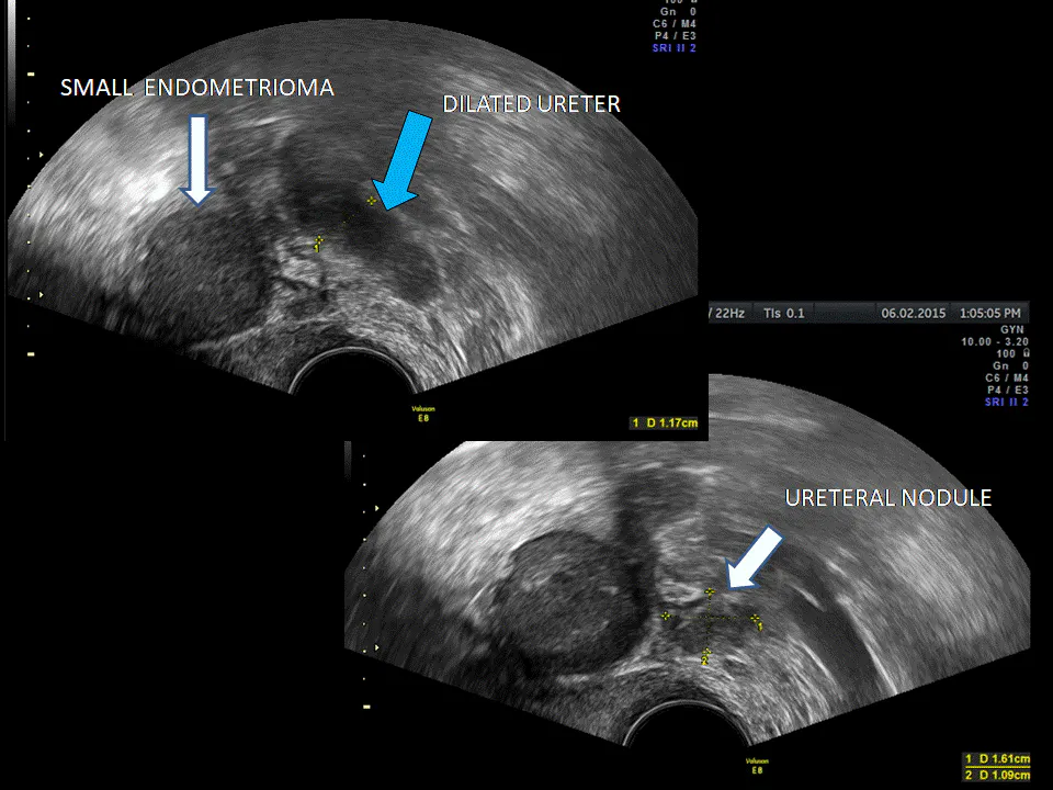
Ultrasound showing ureteral nodule, dilated ureter and endometrioma
Visualizing Ureters With Transvaginal Ultrasound
The systematic nature of a transvaginal ultrasound exam encourages the thorough assessment and documentation of pelvic structures, making it fairly straightforward to add on an examination of both ureters.
For one recommended method for assessing the ureters, begin at the bladder in the sagittal plane by identifying the urethra, then sweep the transducer toward the lateral pelvic wall. The ureter will appear as a hypoechoic tube with a thick, hyperechoic wall. Next, follow the ureter to where it connects to the bladder. Identify and note peristalsis if movement is present, and confirm that the ureter is unobstructed.
Ureters should be assessed based on two factors:
- Presence of nodules: The distal portion of the ureter that can be seen via transvaginal ultrasound extends from the lateral bladder base toward the common iliac vessels. Any focal thickening of the ureter wall or presence of hypoechoic solid nodules can indicate deep infiltrating endometriosis. Nodules must be measured in three planes: length, thickness and transverse diameter. You should also measure the distance from the distal ureteral orifice to the lesion.
- Diameter of the ureter: The average diameter of a ureter at rest is 1.4 to 2.3 mm; during peristalsis, it expands to 2.4 to 3.6 mm. A ureter experiencing stenosis due to endometriosis may be dilated. A measurement of 6 mm or greater taken in the anterior-posterior dimension is considered abnormal and warrants further investigation.
Ultrasound showing dilated ureter and endometriosis nodule (in red)
What If Every Pelvic Ultrasound Included a Bladder and Ureter Assessment?
According to one study, transvaginal ultrasound is a reliable way to diagnose ureteral stenosis in patients with known deep infiltrating endometriosis. Adding ureter assessment to your pelvic ultrasound exams could mean a step forward in endometriosis diagnosis as well as a chance to eliminate years of pain and more invasive procedures for your patients. Try looking for the ureters during your next transvaginal ultrasound — you might just be surprised by what you find.