

Most physicians are familiar with the classic endometrial cancer symptoms and signs — notably, abnormal premenopausal bleeding and postmenopausal bleeding. Fortunately, most women with endometrial cancer are diagnosed early and their survival rates are high. However, patients diagnosed with high-risk nonendometriod cancers, grade III cancers and cancers that have deeply invaded the myometrium have a poorer prognosis.
Endometrial Cancer Ultrasound: A First-Line Tool
There are several different approaches to detecting endometrial cancer. Some clinicians perform a simple biopsy in the office, whereas others perform in-office hysteroscopy. Generally speaking, a point-of-care ultrasound should come first, before any other procedure.
The annual wellness exam is a prime opportunity to review a patient's medical history, discuss gynecologic problems that might arise at their age and offer risk reduction strategies, such as maintaining a normal body mass index (BMI) and regular exercise. A patient questionnaire or review of a menstruation calendar can help you identify patients who may need further evaluation.
A high index of suspicion will serve you well. Recall that 10 percent of women may have abnormal vaginal discharge without bleeding as the presenting symptom. Investigate further for other causes of vaginal discharge if repeated evaluations for cervicitis and vaginitis are negative. Although the average age of diagnosis for endometrial cancer is 65, women younger than 45 are not immune. Some of these women will have a history of polycystic ovary syndrome (PCOS), but some may not exhibit any of the classic risk factors.
Choosing Pelvic Ultrasound Before Biopsy
The days of conducting a blind endometrial biopsy without diagnostic imaging are over. It's best to do a pelvic ultrasound first. This will provide you with information about the endometrial thickness, which is a reliable estimate as to the presence of endometrial cancer and one of several signs of endometrial cancer on ultrasound. Evidence of a thin and regular endometrium (typically less than or equal to 4 mm) can rule out most endometrial cancers, but a blind endometrial biopsy can compromise the ultrasound measurement of the endometrium and render it unreliable.
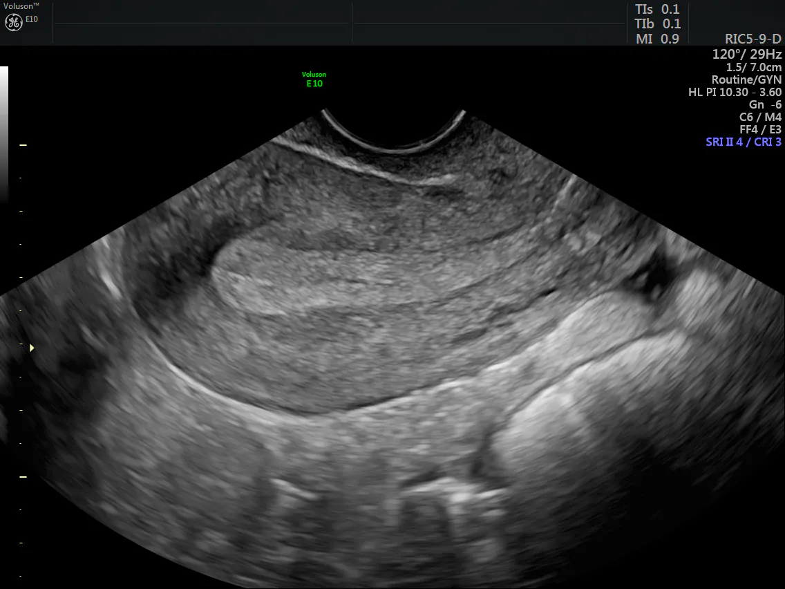
endometrial cancer ultrasound - secretory phase endometrium
New data has shown that the presence of irregular endometrial-myometrial borders, multiple branching vessels and a high color score correlate with the presence of a high-risk cancer. A recent study published in Ultrasound in Obstetrics and Gynecology noted that "grayscale and color Doppler ultrasound features are associated with grade and stage, and differ between high- and low-risk endometrial cancer."
Both endometrial cancer ultrasound in 3D and a saline infusion sonogram (SIS) can help you determine whether there is a focal or global issue. SIS is safe to perform and has not been linked to an increased risk of spreading tumor cells.
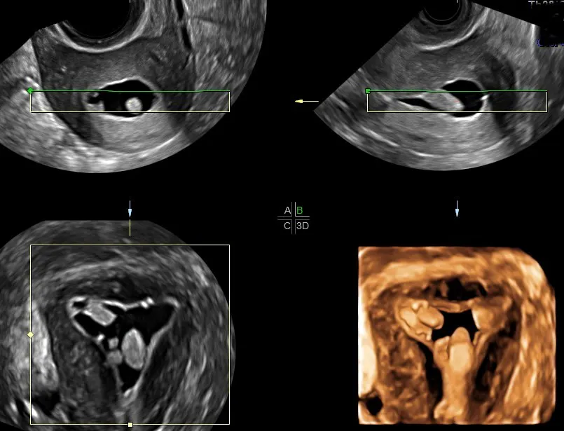
3D Saline Infusion Endometrial Cancer Ultrasound - Uterine Polyps
By pinpointing the area(s) that needs tissue sampling, physicians can more confidently plan a methodical course of action. If the SIS demonstrates a global process, an endometrial biopsy can be performed during the same visit. For a focal disease, an operative hysteroscopy is preferred. A blind office biopsy or dilation and curettage may incompletely remove the lesion or miss it, thus delaying diagnosis. Storage of images and consultation via Tricefy allows for quick confirmation with colleagues while you await final pathology.
Catching Endometrial Cancer Symptoms Early
Endometrial cancer is the most common cancer of the genital tract. The World Cancer Research Fund International estimated that 320,000 cases are diagnosed annually around the globe and that 4 in 10 cases could have been prevented by maintaining a healthy BMI and exercising regularly.
Although there is no single, simple diagnostic screening tool for endometrial cancer, early symptoms typically lead to evaluation and diagnosis. Educating your patients about endometrial cancer symptoms and signs and closely evaluating high-risk populations — combined with rapid, in-office evaluation with ultrasound first — can help improve detection of endometrial cancer and impact outcomes in a positive way.