

Treatment methods for patients with urinary incontinence vary based on the type of incontinence they're experiencing. Diagnosing urinary incontinence with 3D ultrasound offers clear advantages over 2D views, leading to quicker and more accurate diagnoses.
A 2008 report in JAMA found 23.7 percent of women have at least one pelvic floor disorder, and 15.7 percent have urinary incontinence. Pelvic floor disorders increase with age, and they also go up depending on the number of vaginal deliveries the patient has experienced.
2D vs. 3D Ultrasound
When comparing 2D vs 3D ultrasound technologies, 2D ultrasound emits ultrasound waves and records what bounces back, but on in a single plane. The sonographer must capture a series of images of the pelvic floor and view each image separately. A 3D ultrasound creates a sweep of ultrasound waves throughout the field of view, and the ultrasound machine produces a three-dimensional image from the waves that bounce back to the transducer. While a sonographer or physician may have to view multiple 2D images to fully appreciate the pelvic anatomy, a 3D machine can produce an volume that can be manipulated and examined from multiple angles to assist in evaluation of the entire pelvic floor.
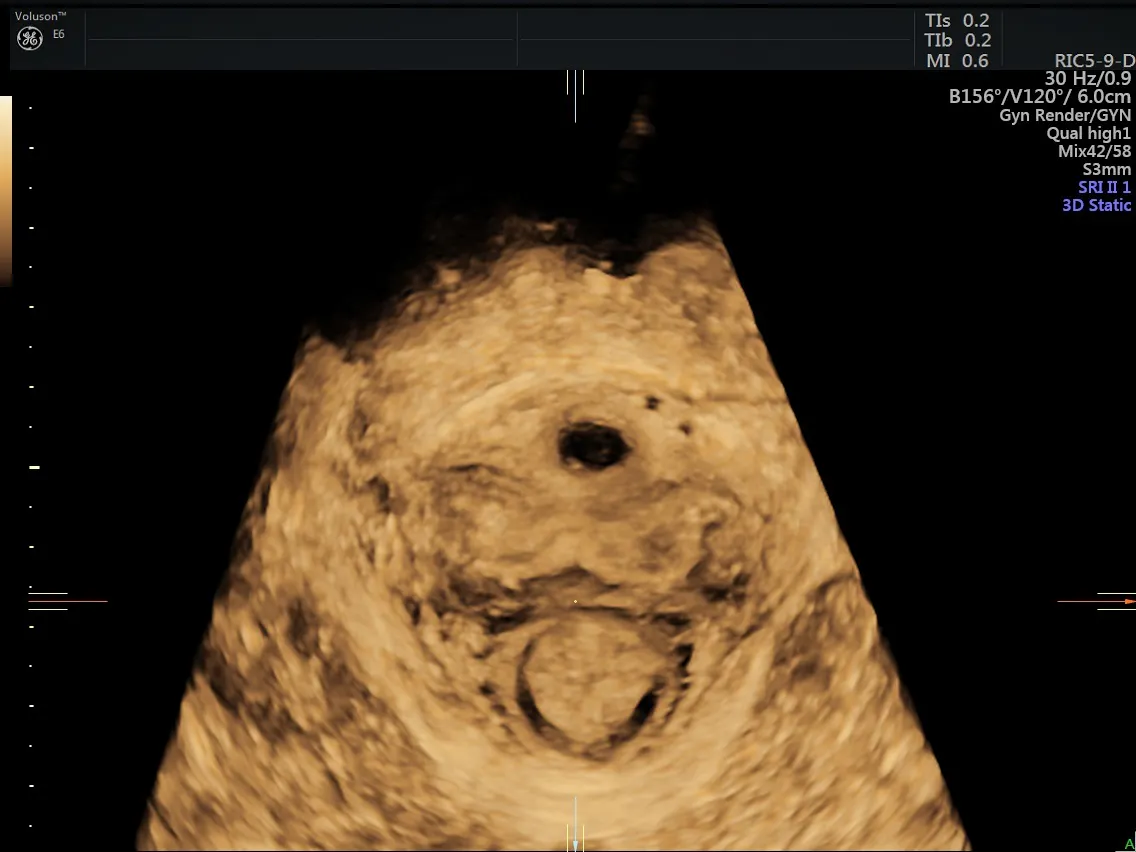
3D Ultrasound image of pelvic floor
Is Diagnosing Urinary Incontinence With 3D Ultrasound Easier?
By using 3D sonography, a physician is able to view the pelvic floor and bladder structures in three dimensions without the time constraints of more costly tests such as MRI. A 2010 article in the journal Ultrasound in Obstetrics and Gynecology states that the advantage of using 3D over 2D sonography to image the pelvic floor and surrounding structures from the transperineal position is the "opportunity to obtain tomographic or multislice imaging" in the axial plane. Sagittal, axial or coronal views may be obtained through transvaginal or endoanal 3D ultrasound imaging. The largest benefit of 3D over 2D ultrasound mentioned by the author is the ability to view the image while rotating, tilting or slicing it to better understand the anatomy, while measuring distance, area, angles and volumes within the image.
Evaluating patients with urinary incontinence with 3D ultrasound enables the physician to visualize the entire pelvic floor area, including the bladder neck, its funnel and mobility, the urethra angle and length and the volume of the bladder.
A 2010 article in the American Journal of Obstetrics and Gynecology stresses the importance of ultrasound in evaluating patients with pelvic floor disorders. Unlike other imaging modes, ultrasound is able to visualize mesh slings and implants. Many doctors refer to the use of 3D ultrasound as a "reproducible" method for visualizing pelvic anatomy, whereas 2D imaging is more user-dependent. In patients who undergo a surgical procedure to correct their incontinence, having 3D images available to compare before and after is crucial to determining future treatments. In fact, Falkert states in Ultrasound in Obstetrics and Gynecology that 3D ultrasound may even be useful in predicting which women will experience pelvic floor disorders such as urinary incontinence when examined shortly after vaginal delivery.


