
Over the last two decades, the use of diagnostic imaging has increased dramatically. There is a race between ultrasound imaging, positron emission tomography (PET)/CT imaging, MRI and X-ray to become the leading imaging modality in the healthcare field.
This boon is being fueled by several factors, including increased demand by patients and clinicians, wider availability of imaging and improvements to technology. These solutions have all led to more accurate diagnoses and better disease management, but some have contributed to ballooning medical costs and increased exposure to ionizing radiation.
Balancing Short-Term Value and Long-Term Risk
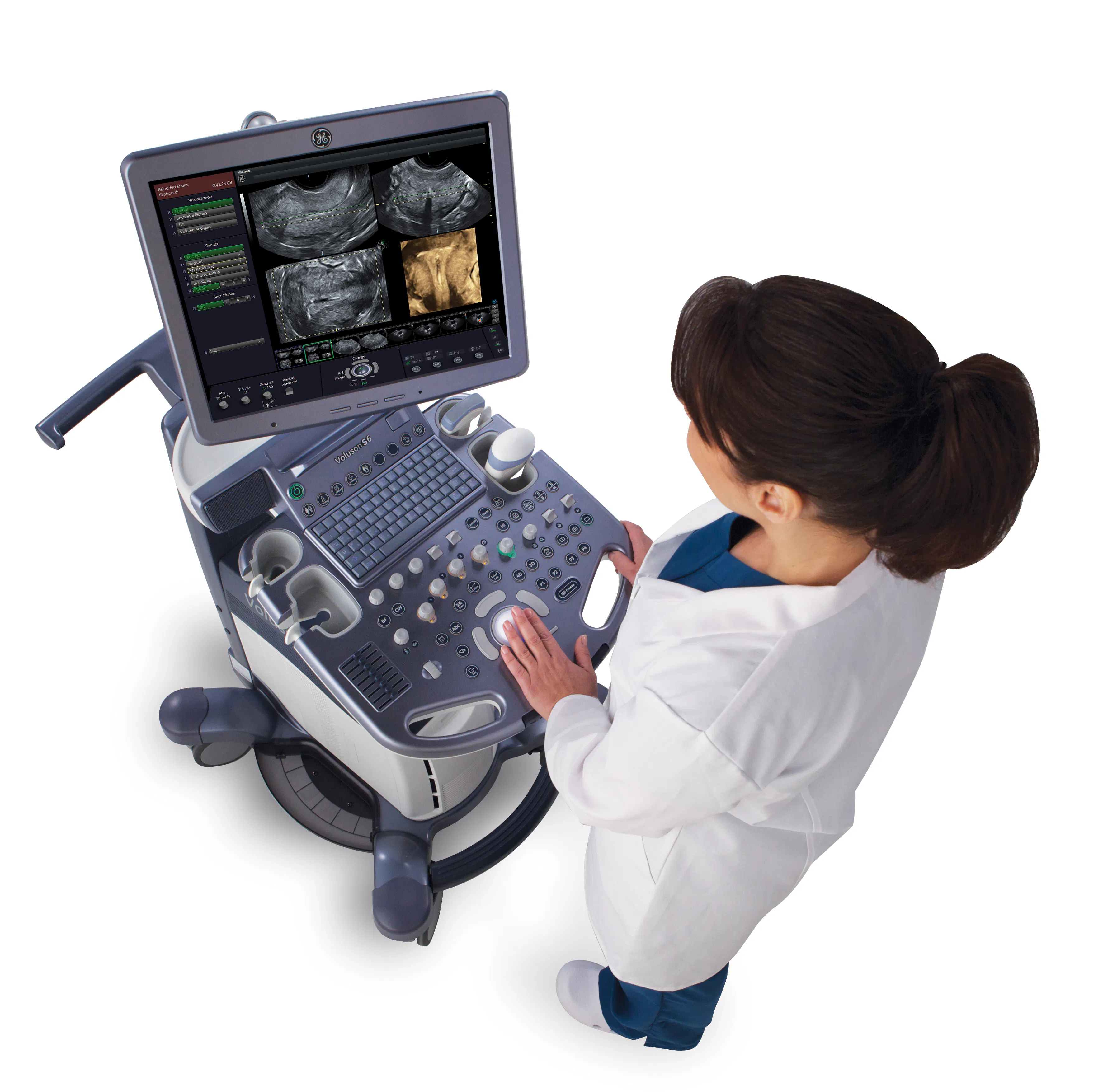
Doctors are coming to see just how valuable and accurate diagnostic imaging tests can be, with nearly every field of medicine using more imaging than they used to. CT and PET scans have gotten faster, X-rays have become sharper, MRI has developed stronger magnets, and ultrasound has become clearer and more powerful. In particular, 3D ultrasound has been a major advancement, providing a coronal plane view that rivals imaging produced by CT and MRI.
However, there is concern about the potential long-term effects of many radiation-based imaging modalities. One potential consequence of increased imaging is substantial radiation exposure from CT, PET and X-ray procedures, which add risk incrementally over a person's lifetime. The U.S. Nuclear Regulatory Commission (NRC) states that any increase in exposure to radiation, including from imaging tests, may increase a person's risk of developing cancer. Ultrasound has been growing and improving without the use of radiation. For this and many other reasons, ultrasound resolution enhancements have rapidly gained ground on more traditional magnetic and radiation-based imaging methods, providing easy-to-interpret results without breaking the bank.
For gynecologists working with reproductive-aged females, the application of any radiation can do more harm than good. This is where advances in ultrasound for gynecologic imaging really start to shine. Ultrasound harnesses the power of high-frequency, nonionizing sound waves and uses Doppler for registering blood flow. This dynamic, interactive technology is portable enough for physicians to offer exams right from the office, provides immediate information for treatment planning and enables online collaboration with cloud-based image sharing.
More Realistic Ultrasound Imaging
3D ultrasound has even more advantages for gynecologists. The one-button feature of an added coronal plane allows for the rendering of 2D slices into a 3D model that more realistically captures an entire organ and its related pathology.
When used in combination with a transvaginal approach, 3D ultrasound allows for digital reconstruction of 3D planar views. According to a study published in the Journal of Obstetrics and Gynaecology of India, this new digital landscape provides almost 100 percent accuracy when determining structural abnormalities of the uterus, such as Müllerian duct anomalies, leiomyomas, endometrial thickening, polyps, ovarian cysts, pelvic adhesions, endometriosis and adenomyosis. Transvaginal ultrasound is also more comfortable for many patients because, unlike transabdominal ultrasound, it doesn't require them to hold a full bladder.
Simplifying Complex Gynecologic Pathology
The list of readily visualized gynecological pathologies and routine assessments grows each year as the field of ultrasound imaging advances. 3D ultrasound assessments allow gynecologists to quickly and easily apply technology to better serve their patients and guide their clinical practices toward success.


