

An adnexal mass is a lesion arising from the ovary, fallopian tube or surrounding connective tissue of the pelvis. It is a common gynecological problem that can be found in females of all ages.
Multiple modalities can be used to assess adnexal masses and determine their origin and type. However, the most successful process currently available relies on adnexal mass ultrasound results to determine a differential diagnosis. Ultrasound's effectiveness stems from its ability to accurately visualize the distinctive features of all adnexal lesions.
Although typically benign, some adnexal masses can be malignant. The main concern is determining if the mass is ovarian cancer, one of the most common cancers in women which also has a poor prognosis.
How the IOTA System Simplifies Adnexal Mass Ultrasound Assessment
A transvaginal ultrasound by an expert practitioner is the best way to determine if an adnexal mass is malignant. Knowing this, the International Ovarian Tumour Analysis (IOTA) group developed rules for assessing adnexal masses.
When done by transvaginal ultrasound the IOTA Simple Rules system describes five features typical for benign tumors (B-features) and five features typical for malignant tumors (M-features). Based on which of the B- and M-features apply, tumors are classified as Benign (only B-features), Malignant (only M-features) or Inconclusive (either no features apply or both B- and M-features appear).
If the B-features can all be applied, cancer can be reliably excluded. If M-features can be applied, the mass is malignant and the patient is referred on to a gynecologic oncologist for staging and treatment. The remaining minority of inconclusive cases call for further assessment by a specialist.
The IOTA Simple Rules can differentiate between malignant and benign in about 80 percent of adnexal masses, and the Asian Pacific Journal of Cancer Prevention applauded this high diagnostic performance.
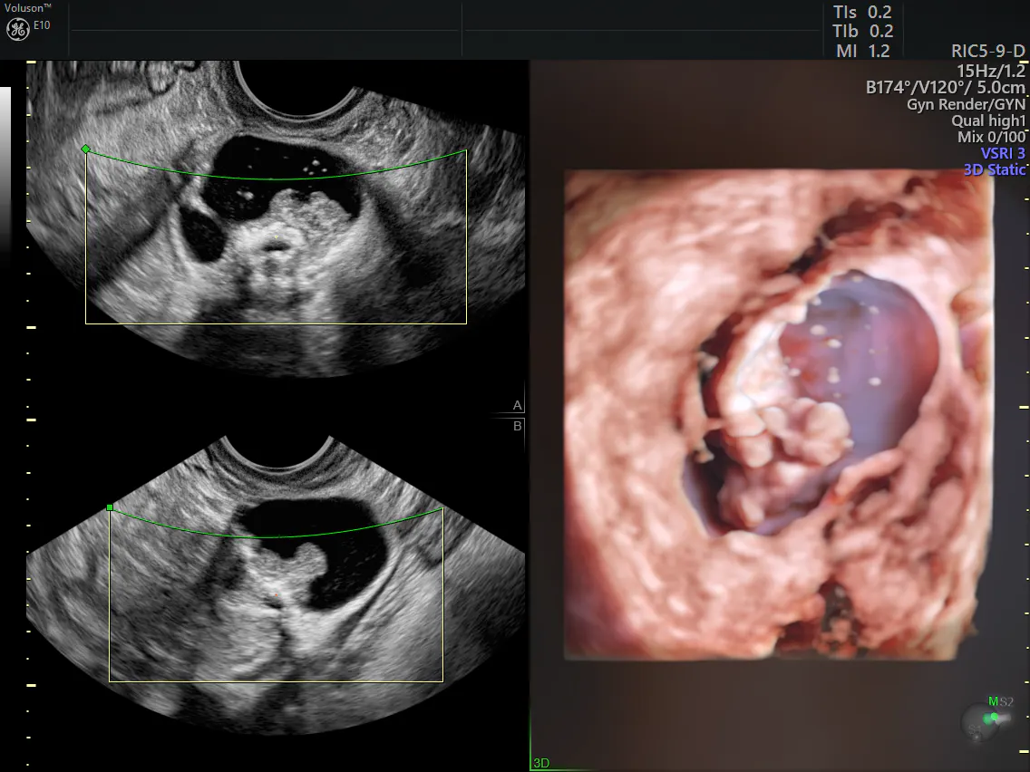
Adnexal mass ultrasound results in 3D
Other Adnexal Mass Differential Diagnosis Systems
Many scoring systems have been developed to increase the ability of practitioners in predicting the malignant potential of an ovarian mass. They focus on objective evaluation of changes in morphological parameters, such as volume, echogenicity, wall thickness, septations, papillae and solid components.
These can be combined with blood testing for serum CA 125 and sometimes other protein markers, such as human chorionic gonadotropin (HCG) and alpha-fetoprotein (AFP), which some germ cell cancers release.
One of the most commonly used alternative scoring systems is the Risk of Malignancy Index (RMI). It includes ultrasound, serum CA 125 levels and the menopausal status of the patient. The ultrasound features of the adnexal mass are entered into a complex model along with menopausal status and CA 125 serum levels to reveal an RMI value that can be interpreted into a high or low risk of malignancy.
Unfortunately, the Journal of Obstetrics and Gynecology has found that serum CA 125 has not been shown to be helpful in determining between benign and malignant ovarian masses leading to the RMI having the poorest performance compared to other methods.
Making Expert Assessment Available to Everyone
The limiting factors for early ovarian cancer diagnosis are both the vague symptoms accompanying the disease and the lack of standardized terms and procedures in gynecological sonography. Early response to ovarian adnexal mass symptoms, identification of ovarian carcinomas and a referral to a specialist are crucial to ensuring accurate staging and optimal treatment.
Recent studies, such as those found in Ultrasound in Obstetrics and Gynecology, reveal that the RMI is not as useful as previously believed in assessing adnexal masses and yielding conclusive results. It is now known that differential diagnosis of ovarian mass or adnexal masses is most conclusively accomplished with an experienced ultrasound practitioner. The IOTA model has found a way to mimic this approach, providing the best way to determine conclusively between benign and malignant ovarian masses without the need for an expert practitioner.
The IOTA Simple Rules uses an ultrasound-based technique that is very useful for clinicians with limited experience scanning ovarian tumors and for teaching purposes. This ultrasound-based system seeks to transfer some of the expert knowledge by offering a clear classification of adnexal masses as either benign or malignant, with the remaining uncertain cases to be referred to an expert practitioner for assessment and diagnosis. With fewer indeterminate cases to address, this results in shorter wait times, faster treatments and a higher survival rate for any patient found to have ovarian cancer.
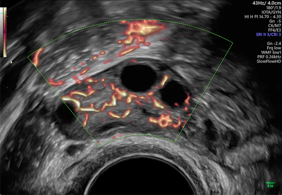
An ultrasound.