

Defined as frequent, heavy menstrual bleeding that may last longer than seven days, menorrhagia can result in pain, lack of vitality, and poor emotional and mental health for the people who experience it. These symptoms often negatively impact their quality of life, according to International Scholarly Research Notices. Menorrhagia can also be a symptom of uterine cancer.
Transvaginal sonography is a primary tool for investigating any type of abnormal vaginal bleeding. However, the causes of this condition can be difficult to pinpoint with menorrhagia ultrasound assessment alone. This is where saline infusion sonography (SIS) and gel infusion sonography (GIS) come in to help clinicians visualize the endometrium and provide a high-quality sonographic assessment.
How Gel Infusion and Saline Infusion Sonography Work
Both SIS and GIS work by slightly distending the walls of the uterine cavity with fluid to visualize the endometrial lining. This is especially helpful if the endometrium is thickened or hyperechoic. Both techniques are ideally performed before day 10 of the menstrual cycle, when the endometrium is thinnest. Neither procedure typically requires anesthesia.
In both SIS and GIS, a catheter is inserted through the cervix before a transvaginal probe is inserted into the vaginal canal and placed within one of the cervical fornices. Warmed sterile saline solution or gel is then slowly instilled into the endometrial canal while a clinician performs the ultrasound exam.
What GIS, SIS and Menorrhagia Ultrasound Reveal
Endometrial polyps, endometrial hyperplasia, intracavitary fibroids or lesions, and endometrial cancer can all cause menorrhagia; all of them can be revealed by SIS and GIS. Infusion sonography can also help clinicians discover endometrial atrophy, adhesions, congenital defects of the uterus and signs of focal adenomyosis.
Doppler ultrasound can be used alongside fluid infusion. It allows visualization of blood flow to see and evaluate blockages (such as clots), the flow stalk in polyps, the peripheral flow of fibroids and the disorganized vascularity of tumors.
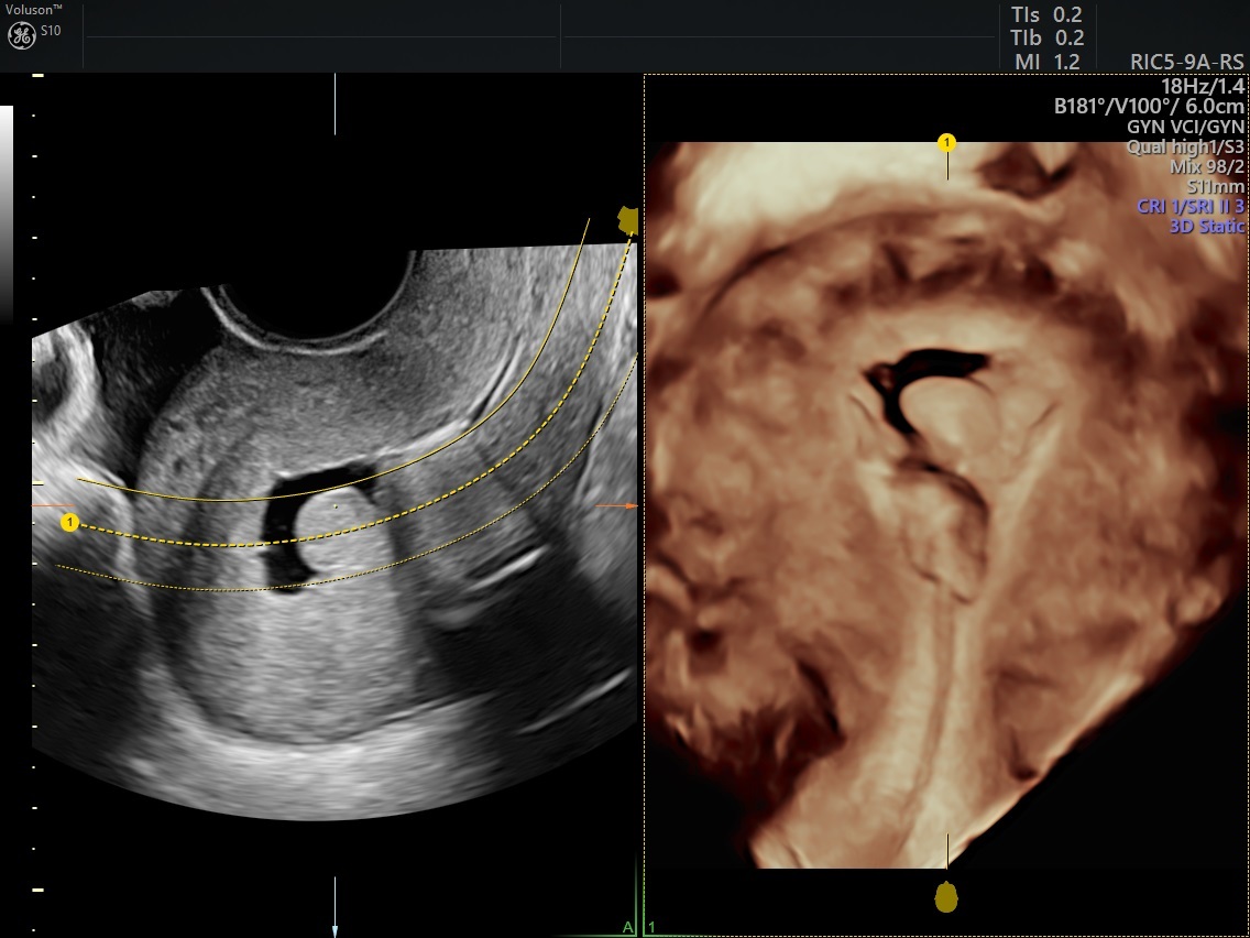
SIS with polyp for menorrhagia ultrasound
The Difference Between GIS and SIS
Ultrasound in Obstetrics and Gynecology reports that SIS is a highly sensitive method for detecting endometrial polyps and submucosal uterine fibroids, making it an effective primary diagnostic tool for assessing abnormal uterine bleeding.
However, the saline infusion sonography procedure was found to frequently cause either imaging failure (when saline back-flowed out of the uterus) or significant patient pain, depending on the catheter used.
Gel infusion sonography was developed to fill the uterine cavity more stably. It uses the same infusion technique with a higher viscosity gel in place of saline. The Middle East Fertility Society Journal reported that the advantages of GIS allowed for longer investigation time and was more sensitive than SIS. Fertility and Sterility found that GIS is a feasible, accurate alternative to saline with fewer technical failures.
However, in a much smaller study published by Obstetrics and Gynecology, the image quality of saline was found to be slightly superior, likely due to the presence of air bubbles in the gel. The gel was able to distend the uterine cavity slightly more than saline and hold a more stable distension for at least four minutes, making it a suitable alternative to saline as long as air bubbles can be prevented.
Adding Fluid Infusion to Your Practice Repertoire
Both SIS and GIS have shown remarkable results in identifying the causes of menorrhagia. This benefits both patients and clinicians; the procedure is less invasive than a hysteroscopy, yet more accurate than the average pelvic ultrasound, minimizing the need for analgesics and allowing for quick and effective treatment of abnormal or heavy uterine bleeding. Fluid infusion is a smart addition to the ways you already use menorrhagia ultrasound to provide a comfortable and effective treatment for your patients.
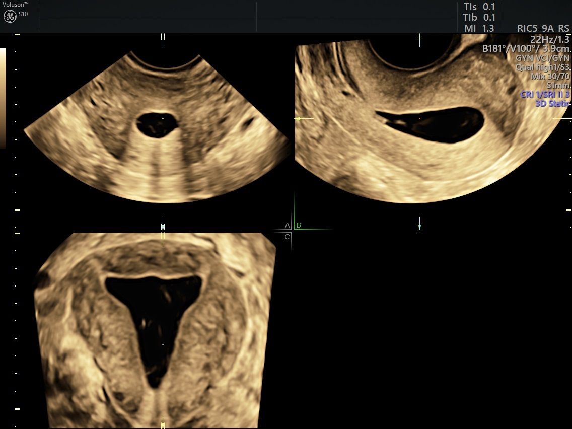
SIS multiplanar imaging for menorrhagia ultrasound
Disclaimer: HyCoSy is not cleared in the United States and may not be available in all other countries.
Read more about this topic in the case study: "The Standardized Approach to Endometriosis Screening."

