
As technology becomes smaller, faster, smarter and more user-friendly, many areas of medical imaging have seen major improvements. Ultrasound imaging, specifically, has seen advancements in usability, image resolution enhancement and cost-effectiveness, as well as a widespread increase in application. These developments can help gynecologists reduce misdiagnoses and build trust with patients.
3D ultrasound is easy to learn, and provides clarity and certainty in the diagnosis of complex gynecological issues. This enables clinicians to provide timely, effective treatment for their patients.
The Evolution of 3D Ultrasound Imaging Technology
Recent advancements in ultrasound image resolution of both objective and theoretical origins have shown a great deal of potential. In the past, the focus was on improving post-processing image resolution, but recent research has focused on automatic resolution improvements that work with both image acquisition and post-processing features.
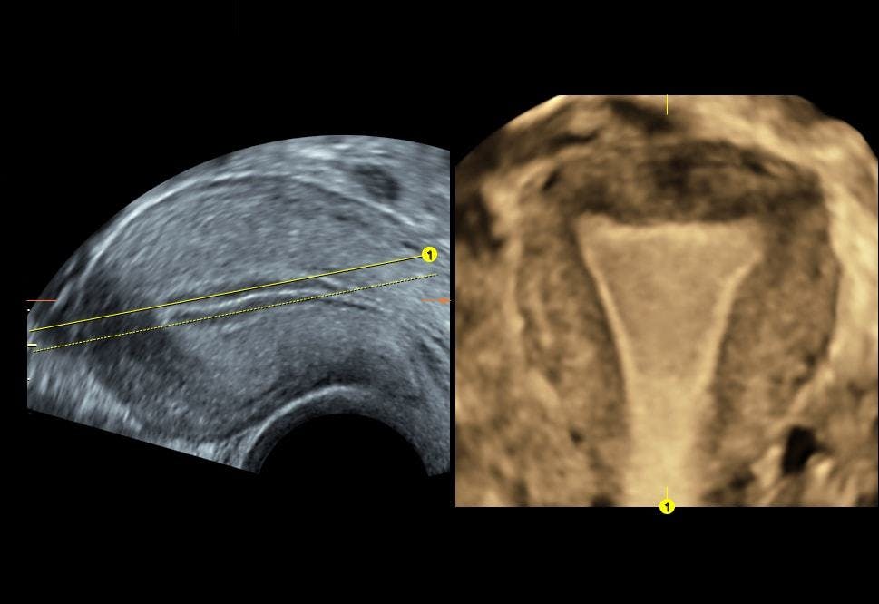
Take the diagnosis of a septate uterus, the most common structural abnormality of all Müllerian duct defects. The presence of an intrauterine septum can lead to miscarriage, preterm labor or recurrent in vitro fertilization (IVF) failures, making this an important diagnosis for the patient. Traditionally diagnosed with more invasive methods such as hysterosalpingography and hysteroscopy, a septate uterus can now be more accurately diagnosed with 3D transvaginal ultrasound, according to research published in the Journal of Obstetrics and Gynecology of India. Transvaginal ultrasound imaging is less invasive, quicker and can be done right in the gynecologist's office, allowing for immediate diagnosis and treatment planning.
The advantage of 3D transvaginal ultrasound is the ability to produce a coronal view of the entire fundal surface of the uterus, as well as multiple planes through the endometrial canal. This enables clinicians to precisely evaluate fundal indentation and septum length, minimizing misdiagnoses.
Improved Accuracy and Volume Calculation
As for the rest of the spectrum of congenital Müllerian duct anomalies, 3D ultrasound can also accurately differentiate between the classes of unicornuate, bicornuate, uterus didelphys, septate uterus and arcuate uterus. The aforementioned study in the Journal of Obstetrics and Gynecology of India found the sensitivity and specificity of 3D transvaginal ultrasound to be close to 100 percent, which is comparable to MRI and laparoscopy. Even in the cases of more common uterine lesions such as fibroids, adenomyomatosis and intrauterine synechia, 3D ultrasound has been found to be more accurate in defining borders and accurate measurements.
Part of the improvement in gynecological 3D ultrasound is due to a new application known as automated volume calculation, which identifies hypoechoic structures and calculates their volume from the acquired 3D volume dataset. This application can reduce examination time, make accurate measurements and be highly reproducible.
Building Trust Through Technological Advancement
It is clear to see that adding 3D ultrasound to a routine gynecological workflow can be beneficial for clinicians looking to provide timely and accurate results in a relatively easy and cost-effective manner. The ability to provide the patient with quick, accurate and inexpensive diagnosis during her visit builds a strong base of trust when it comes to both diagnosis and efficiency of treatment.


