
When it comes to diseases commonly affecting women, endometriosis isn't typically the first that comes to mind. But this condition is more common than many may think.
More than 11 percent of women in the U.S. between the ages of 15 and 44 may have endometriosis, according to the U.S. Department of Health and Human Services' Office on Women's Health. It is a common cause of pelvic pain for women in their 30s and 40s.
When determining how to detect endometriosis, laparoscopy is the gold standard — but 3D ultrasound can add valuable information before surgery, especially in diagnosing and evaluating deep infiltrating endometriosis.
Evaluating Endometriosis
Endometriosis is a condition in which tissue similar to the type that normally grows inside the uterus grows outside of it. This tissue can appear on the bladder, ovaries, bowels or fallopian tubes. Tissue can penetrate beneath the peritoneal layer, leading to a condition called deep infiltrating endometriosis, a rare form of the disease. The exact cause isn't understood, but it's thought to be driven by inflammation that depends on estrogen. Some cellular defects and mutations may also contirbute to the pathogenesis of endometriosis.
Women who suffer from this condition present with pain in the abdomen, lower back or pelvis. It can also cause:
- Infertility;
- Heavy periods or bleeding between periods;
- Painful menstrual cramps;
- Gastrointestinal symptoms;
- Pain during sex;
- Blood in urine; and
- Abdominal bloating.
When the endometriosis spreads to the cul-de-sac, the area between the upper vagina and rectum, it can lead to the rectum and vagina fusing.
When evaluating a patient for suspected endometriosis, use laparoscopy to take a sample of the tissue and test it. Laparoscopy is the only way to develop a definitive diagnosis. However, imaging tests are often used before surgery to evaluate and locate the lesions and ensure the right patients are being sent for surgery. Transvaginal ultrasound can detect endometriomas within the ovary as well as deep infiltrating endometriosis.
Why and How to Detect Endometriosis With Ultrasound
Transvaginal 3D ultrasound is a valuable but often underutilized tool in gynecology. Research has shown it is sensitive and specific for detecting endometriosis beyond the ovaries, according to a review in the Journal of Ultrasound in Medicine.
The authors recommended using transvaginal ultrasound to evaluate the rectosigmoid for bowel lesions, a severe form of deep infiltrating endometriosis. These lesions have a distinct comet shape that can be spotted with an ultrasound. Surgery for bowel lesions may be challenging, but the ultrasound evaluation can help inform surgical decisions.
Although an MRI is commonly ordered to detect deep infiltrating endometriosis, ultrasound has comparable sensitivity and specificity for lesions in the rectum and lower sigmoid colon, at a lower cost to boot. Ultrasound is also a more comfortable procedure for many women and can eliminate the need to travel for additional appointments.
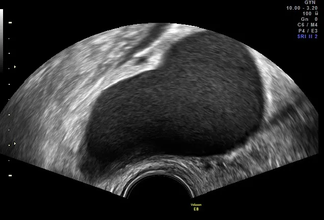
2D ultrasound image
2D ultrasound can visualize nodules or adhesions indicative of deep infiltrating endometriosis.
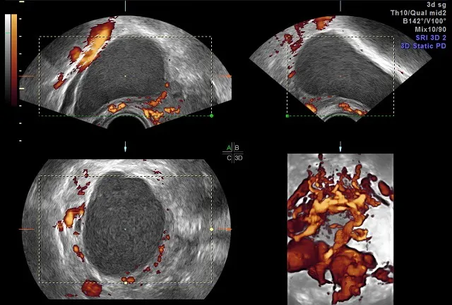
3D ultrasound image
3D ultrasound visualizes endometriomas for a presumptive diagnosis and presurgical planning.
Images courtesy of Professor Stefano Guerriero
Transvaginal ultrasound offers additional benefits in evaluating endometriosis over MRI. A review in specifically for detecting lesions outside the ovaries, which can cause organs to stick together. A review in Diagnostics (Basel) found that transvaginal ultrasound should be used first when the physician suspects deep infiltrative endometriosis. This method is more accessible and less expensive than MRI, making it an essential tool for diagnosis and presurgical planning.
Endometriosis in the bowel or gastrointestinal tract is most often treated with surgery. Detecting these lesions is critical in surgical planning, and performing a 3D ultrasound first adds valuable information to treatment approaches. When planning surgery, ultrasound may detect lesions not visible during laparoscopy. In a case study, published in the Journal of Obstetrics and Gynaecology Canada, a preoperative ultrasound detected deep endometriosis at the left uterosacral ligament. This abnormality wasn't visible during surgery, and ultrasound ensured the surgeon could excise the lesion.
Practices that employ the right tools to glean these insights are well-equipped to provide the right level of care and treatment to address each patient's needs.