How Ultrasound Helps Diagnose Common Pelvic Floor Concerns
The prevalence of pelvic floor disorders (PFD) is astonishingly high. Research published in the Global Library of Women's Medicine reports that as many as 46 percent of women experience at least one form of PFD, such as urinary incontinence, fecal incontinence or pelvic organ prolapse.
But urogynecologic medicine has advanced, and those patients now stand to have better outcomes thanks to innovations in diagnostic and interventional technology. Among those innovations is the modern-day use of 3D ultrasound, which facilitates reproductive health exams with increased visual clarity, speed and ease of use.
Diagnosing Common Pelvic Floor Disorders
Pelvic disorders encompass a wide range of conditions; besides pelvic organ prolapse and fecal and urinary incontinence, they can include perineum trauma from childbirth.
A similarly large number of factors may raise a patient's risk for these disorders, including aging, vaginal childbirth (especially of an infant over 8.5 pounds) and obesity. Depending on the specific disorder, symptoms may include pain, pressure, vaginal bulging, sexual dysfunction and incontinence.
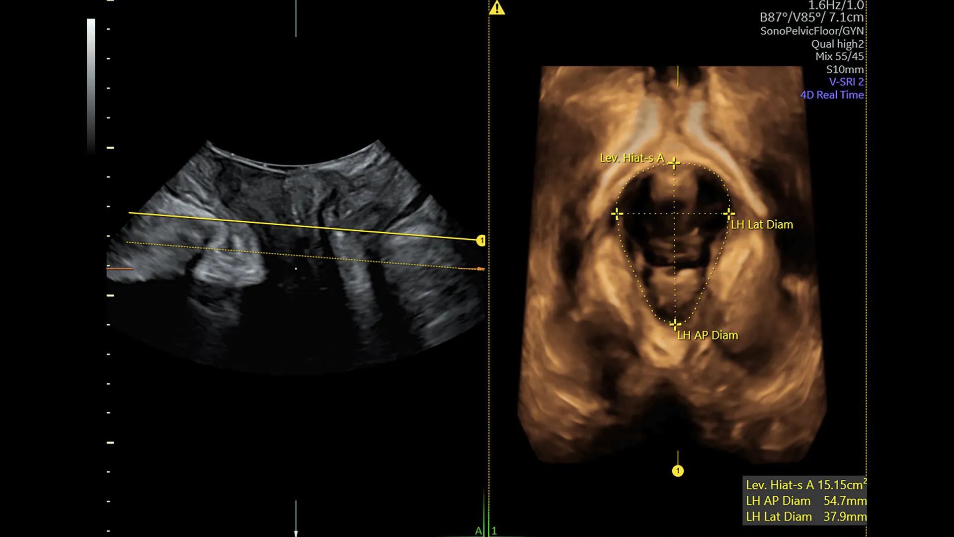
If a pelvic floor disorder is suspected, advanced imaging can help clinicians visually diagnose issues within the pelvic floor. This is commonly done with perineal ultrasound, which allows non-invasive imaging without distortion and is available in either 2D or 3D.
However, compared with 2D ultrasound, which visualizes only the midsagittal and coronal planes, 3D ultrasound adds additional clinical utility by incorporating the axial plane. This helps further discern and diagnose a number of complications related to pelvic conditions.
Diagnosing Anterior Prolapse
Descent of the anterior vaginal wall is commonly due to — and blanket-labeled as — a cystocele. However, as one paper in Current Surgery Reports remarks, that may not be the sole reason behind prolapse in the anterior compartment. The root cause may lie with other underlying issues, such as urethral diverticulum, anterior enterocele and Gartner duct cyst.
Discerning between these many anterior conditions requires ultrasound imaging, which can be rendered more useful with a 3D platform. In particular, this guidance may be helpful:
A cystocele is bladder prolapse. It can be assessed by comparing viewpoints of a patient at rest with the patient performing the Valsalva maneuver. Add a horizontal line on the screen that goes through the inferior margin of the pubis symphysis or, if using a touch screen, draw one. The distance that the bladder descends indicates the extent of the cystocele and helps to quantify the grading of descent from mild to moderate to severe.
Urethral diverticulum describes a sac formation along the urethral wall. It is particularly discernible on the axial plane, which depicts the urethral anatomy in a delineated manner. Look for an anechoic representation, similar to the bladder; it generally manifests posterolaterally on the urethra's midportion. This condition may be overlooked as a culprit in patients with recurring urinary tract infections or complaints of urinary pain, frequency or urgency.
Anterior enterocele is the prolapse of the small intestines. It can be seen anteriorly as abdominal contents that descend down, but it is more commonly seen posteriorly in older patients after a hysterectomy or other procedures.
Gartner duct cysts are remnants of wolffian ducts. They typically only cause symptoms when enlarged or infected. These cysts are particularly discernible on the axial plane. However, unlike urethral diverticulum, they will not be connected to the urethra. Look on the upper anterolateral wall of the vagina, above the inferior part of the pubic symphysis. The cysts appear anechoic (or echogenic, if infected).
Alternatives to perineal ultrasound such as lateral urethrocystogram and fluoroscopic imaging can also help diagnose anterior conditions. Compared to these alternatives, perineal imaging has been shown to be as effective — and sometimes better — at both diagnosing prolapse conditions and quantifying the extent of damage before and after surgery (such as by assessing bladder neck condition or mobility).
Diagnosing Posterior Prolapse
Similarly to anterior prolapse, Current Surgery Reports authors also point out another term, rectocele, that mistakenly gets canvassed across the entirety of issues involving posterior descent of the vaginal wall. And, as with anterior prolapse, advanced imaging modalities can help specify between these varying pathologies:
Rectocele is rectal prolapse. On ultrasound, it can be detected by a defect in the rectovaginal septum during the Valsalva maneuver and quantified by the extent of the rectum's descent.
Perineal hypermobility is similar to a rectocele in that the rectum inferior is displaced in relation to the pubis symphysis but occurs when the septum is intact.
Posterior enterocele is a small intestine prolapse. It can be seen as an abdominal matter that extends forward to the anorectal muscularis. Unlike rectoceles, which are composed of air and feces, enteroceles will be more homogenous on ultrasound. Rectoenterocele is the dual occurrence of rectal and small intestine prolapse.
Rectal intussusception is thought to be early-stage rectal prolapse due to trauma of the levator ani. It can be identified as a double hypoechoic ring around the rectum, when the rectum folds into the proximal anal canal, as a patient performs the Valsalva maneuver.
Tears from childbirth are repaired after delivery, but lasting external anal sphincter damage can occur; for some patients, this damage results in lasting symptoms, such as fecal incontinence. Because these tears can lead to pelvic organ prolapse and incontinence, clinicians should be aware of these defects and their predisposition to other pelvic floor disorders.
In addition to ultrasound, other available diagnostic techniques (employed by colorectal surgeons) include defecation proctography — however, this procedure can be uncomfortable and involves radiation exposure. It also does not offer diagnostic value elsewhere in the levator ani, which is a limitation given that other problems can coexist with posterior vaginal wall descent.
Supporting Diagnostic and Interventional Decision-Making
Even after the initial diagnosis of a pelvic floor disorder, ultrasound continues to play a role as an aid to pelvic reconstructive surgery, as it can help physicians identify synthetic implants, tapes, slings and postsurgical complications.
3D ultrasound gives a more expansive and anatomically comprehensive scan of the female pelvis, but it is not just the dimensional perspective that supports these diagnostic and interventional decisions. Machines with ease-of-use features, such as touch screen capabilities or programmable configurations, can also add value and speed to the point of care. By helping make visits as efficient as possible, these features can also make all the difference in patient satisfaction.
Interested in learning more about ultrasound and Pelvic Floor?
Dr. J. Schwayder’s lecture: How to evaluate a patient presenting with Pelvic Pain
Dr. A. Youssef’s lecture: 3D ultrasound of the pelvic floor.
Dr. A. Youssef’s lecture: Pelvic floor ultrasound for obstetric injuries
Dr. R. Kamal’s lecture: Transperineal ultrasound and pelvic floor disorders
Short article: Assessing the pelvic pain patient in 5 easy steps.

