
For many decades, clinicians in the field of urogynecology only had access to the limited imaging data of static cystourethrography, an expensive option that often produced poor-quality assessments of the pelvic floor and related organs.
Pelvic MRI was more effective, but due to high cost, slow acquisition speeds and inaccessibility, it was rarely used clinically. The recent increased availability of ultrasound, with its dynamic imaging, low cost and wide availability, has renewed interest in imaging within the urogynecology and female urology fields, particularly with the use of 3D ultrasound in pelvic reconstruction.
Pelvic Floor Dysfunction and Types of Imaging
Urogynecology treats dysfunction of the pelvic floor, including urinary incontinence, pelvic organ prolapse and fecal incontinence, as well as trauma to the perineum experienced during childbirth. According to the National Institute of Health, pelvic floor disorders affect nearly one in four women in the United States, and of those, 10 to 20 percent will require surgery due to organ prolapse.
Assessment of the pelvic floor to address these issues has been done with transabdominal, perineal, transrectal and transvaginal ultrasound. Perineal, or translabial, is currently the most widely used due to its noninvasive nature, wide availability and absence of distortion.
Perineal imaging of the lower urinary tract yields information equivalent or superior to the lateral urethrocystogram or fluoroscopic imaging. Ultrasound can expose uterovaginal prolapse and reliably assess bladder neck position and mobility.
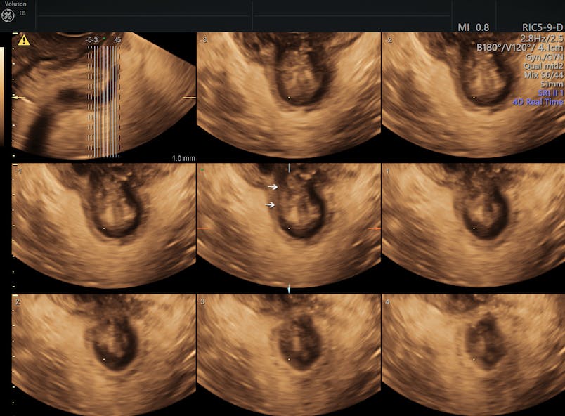
Due to the automatic image acquisition of 3D ultrasound, a multitude of sectional planes, including the midsagittal, coronal and axial planes, can be integrated into a volume using this technology. Access to the axial plane is one of the main advantages of 3D ultrasound, as such access was previously only available by MRI.
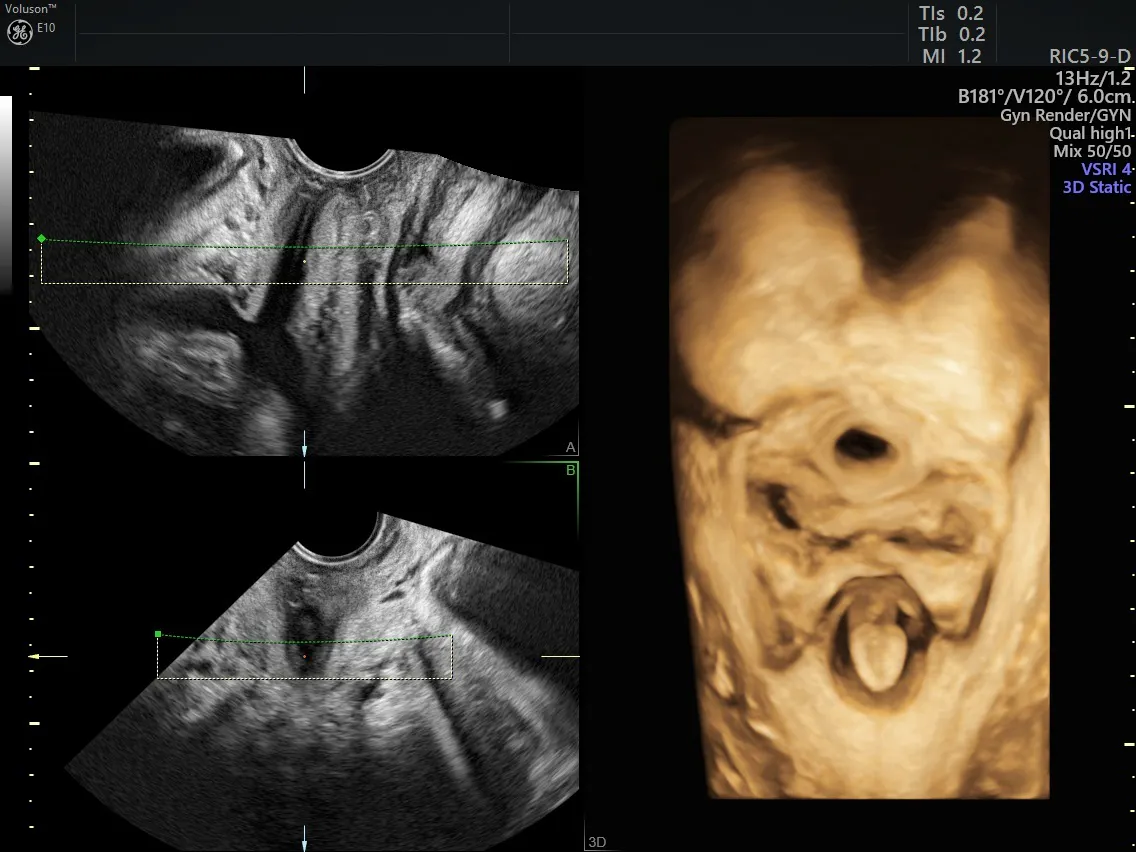
3D Ultrasound Pelvic Floor
Using 3D Ultrasound in Pelvic Reconstruction
A number of pelvic floor issues simply cannot be properly assessed without 3D ultrasound. The technology allows physicians to accurately identify the location of synthetic implants, tape and slings, as well as related complications. It also helps them differentiate between a cystocele, urethral diverticulum, Gartner duct cyst or anterior enterocele in the anterior pelvic compartment.
Today, a truly functional anatomical assessment of the female pelvis is only feasible with 3D volume ultrasound. This encompasses the observation of downwards displacement of pelvic organs on Valsalva, the effect of a contraction and Valsalva on the levator hiatus, the development of prolapse and the opening up of fascial or muscular defects.
Convenience and Simplicity
The convenience with which medical professionals can now obtain pre‐ and post‐treatment imaging data will simplify outcome studies after prolapse and incontinence surgery, and it will enable the development of improved surgical procedures, individualized reconstructive surgery and early detection of complications.


