
If you've ever referred a patient for ultrasound imaging tests, you already understand the benefits of this technology in gynecology. Ultrasound allows clinicians to visualize the pelvic anatomy and can aid in the diagnosis of many anomalies. Sending patients to a local imaging suite can uncover and confirm various health concerns, but you can accomplish all that and more for your patients — and your practice — with on-site 3D sonography.
The Benefits of 3D Ultrasound in Gynecology Practices
With the ultimate goal of delivering exceptional care, you want to do everything possible to provide accurate diagnoses and timely treatment. Offering comprehensive ultrasound imaging directly to patients provides your practice with a number of advantages.
- More accurate diagnosis: Sonography has been shown to improve the accuracy of diagnoses in many fields of medicine, including gynecology. With ultrasound right at your fingertips, you can make quick, informed clinical decisions about your patients' health and prescribe treatment with greater precision.
- Increased revenue: On-site ultrasounds can increase value for your gynecology practice, too. Reimbursement from insurers that would otherwise go to outside services will now go directly to you and your practice.
- Cost savings: Conducting ultrasounds within your office can save patients the cost of travel and lost time off work, in addition to avoiding co-pays at other offices or appointments.
- Improved patient satisfaction: In-house ultrasound enables patients to undergo imaging tests in the familiar comfort of your office, administered by a clinician whom they already know and trust.
Medical Advantages of 3D Ultrasound
3D ultrasounds can provide even more benefits to you and your patients. This technology allows gynecologists to view the coronal plane, creating 3D visualizations that more realistically represent the internal organs.
A paper titled "Role of Three-Dimensional Ultrasound in Gynecology" suggested that the use of 3D ultrasound in gynecology can help physicians better identify and diagnose a range of maladies. Below are just a few of the pelvic and gynecologic conditions that are more easily detected using 3D ultrasounds.
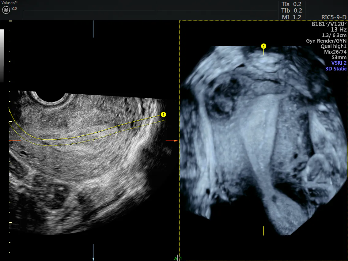
3D Ultrasound image of the uterus and a leiomyoma
Sagittal and Coronal view of Uterus and endometrium. Coronal view shows hypoechoic heterogeneous focal lesion within myometrium, possibly a fibroid.
- Cancer: Ultrasound is typically the initial imaging tool used to evaluate patients with abnormal masses or bleeding, symptoms that can signal gynecological cancer. One review published in Gynecologic Oncology concluded that 3D ultrasound is better suited to detect suspicious adnexal masses than 2D technology.
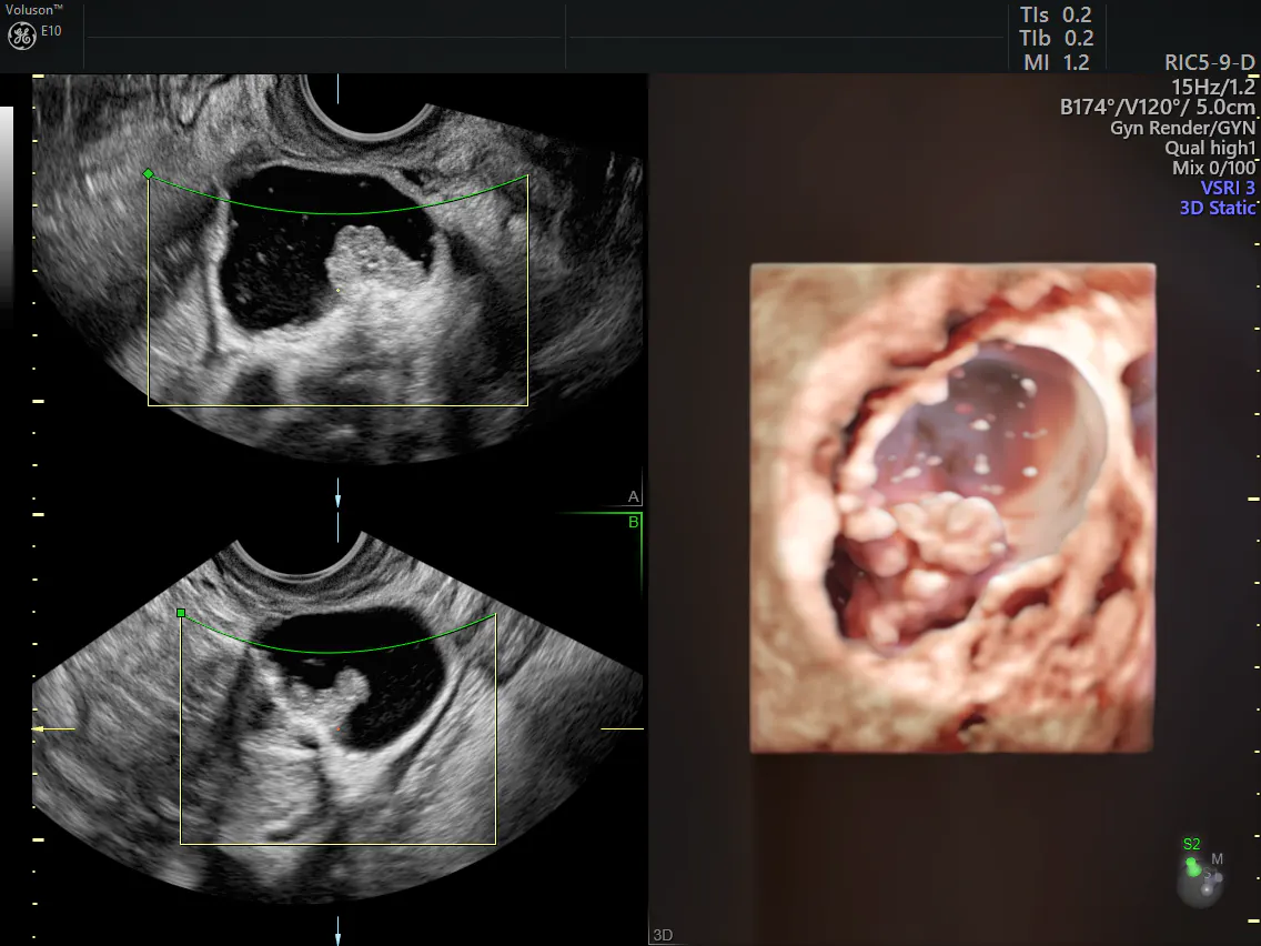
3D image of a complex ovarian mass
Sagittal and Transverse planes of an ovarian lesion (mostly cystic with solid component). These two images were then used to create a 3D view of the abnormality in the 3rd image on the right.
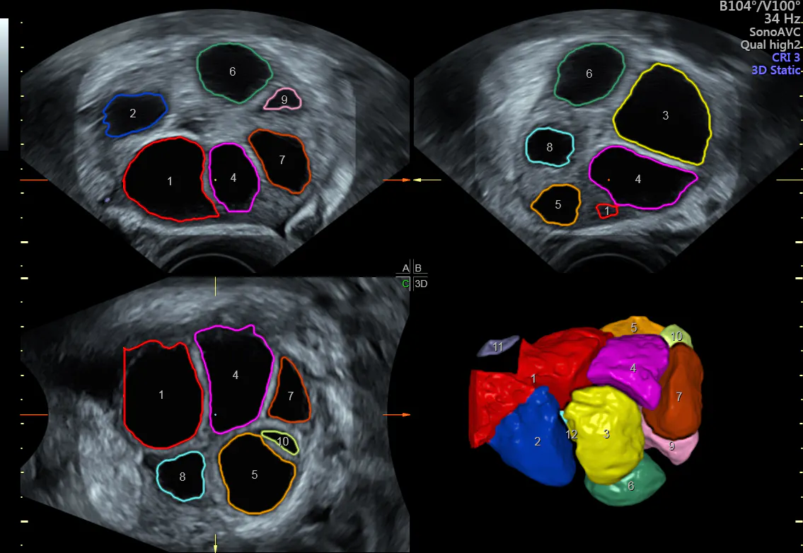
SonoAVC follicle measuring ovarian follicles
Sagittal and transverse planes of an ovary with multiple enlarged follicles present. Each follicle was traced for a size/volume measurement. A 3D color view of the follicles was created and labeled for easy tracking of the number of follicles. Likely for oocyte harvest/retrieval.
3D ultrasound is an indispensable tool for gynecology practices. Given all this empirical evidence, as well as the numerous advantages for patients and practices alike, it's no surprise that a growing number of clinicians are making the switch from 2D to 3D.


