
2D ultrasound is crucial for gynecology assessments, but OB/GYN care can be supported even further with the use of 3D ultrasound. 3D ultrasound is often under-utilized in gynecological cases, where it has the ability to add useful information and allow for improved patient care. A 3D ultrasound image can be critical for assessing endometrial polyps, adenomyosis, uterine fibroids, and congenital uterine abnormalities. 3D ultrasound is also instrumental in placing and monitoring intrauterine devices (IUDs) and help to find some causes of infertility.
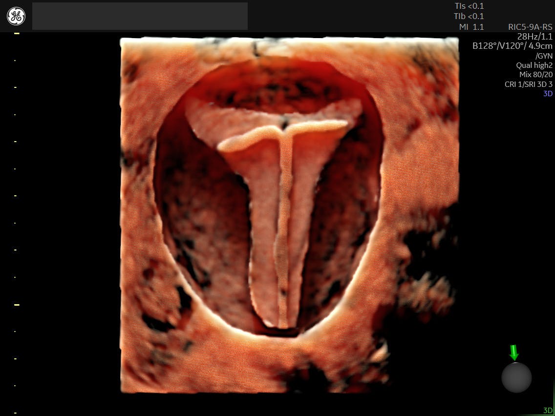
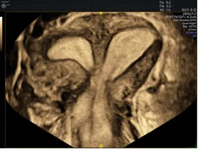
3D Coronal View, Partial Septate Uterus
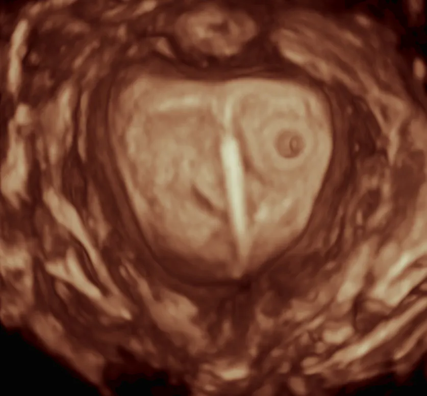
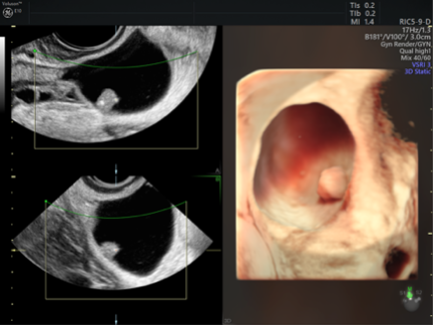
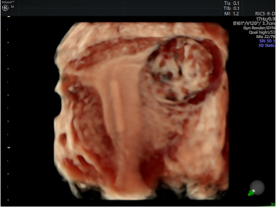
Rendered 3D coronal view showing IUD in early pregnancy.
To ensure a successful 3D ultrasound exam, physicians should have an approximate idea of the dimensions of the uterus or other organ being examined. Acquiring an accurate 3D ultrasound volume requires knowledge of volume acquisition, volume display and volume analysis.
Volume Acquisition
For a typical uterine sweep, open the sweep wide (120°) to include the cornua. Smaller volume angle can be used for a faster acquisition if needed. For an ovarian sweep, the volume angle can also be smaller, ensuring the sweep begins just before the ovary is visualized and ends just after the ovary.
The sweep time is dependent upon the Quality setting - the higher the Quality, the slower the acquisition speed. Using a higher quality will result in higher resolution of the volume. If background artifacts are present, they can be minimized by reducing the acquisition time.
Volume Display
Below are three commonly used 3D display formats. Understanding the differences between them will in the selection of the most beneficial format for the anatomy you are investigating.
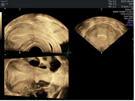
1. Multiplanar Reconstruction (MPR). MPR simultaneously displays 3 orthogonal planes, typically sagittal, transverse and coronal (these would be A, B and C) planes. The reconstructed 3D rendered image can not only be optionally displayed, but can also be shown as a single display, without the A, B and C planes.
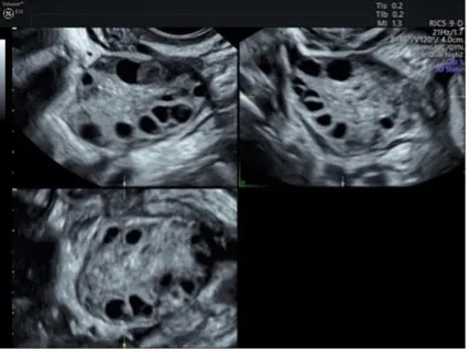
Polycystic ovary shown using 3D ultrasound
Gynecological cyst shown using 3D ultrasound, rendered with HDlive™Studio
Caesarean Section Scar shown using 3D ultrasound
2. Tomographic Ultrasound Imaging (TUI). A multi-view format that demonstrates sequential slices through a volume of data for a CT or MRI-like appearance. It is especially useful in assessing pathology of the pelvic adnexa, such as ovarian and paraovarian cysts, as well as fallopian tube abnormalities.
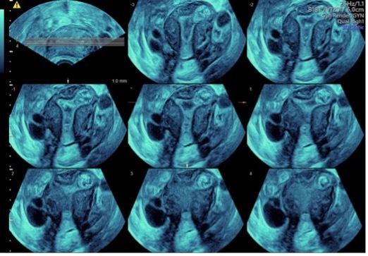
3D uterus in TUI display, acquired with Voluson™ SWIFT
3. Speckle Reduction Imaging (SRI). Speckle Reduction Imaging is an imaging algorithm that improves the image quality of B-mode scanning by reducing speckle for a more tissue-like appearance.
3D coronal plane helps differentiate between these uterine anomalies. SRI reduces speckle for a more tissue-like appearance, and can enhance visualization of cystic masses ,endometriosis, adenomyosis and more.
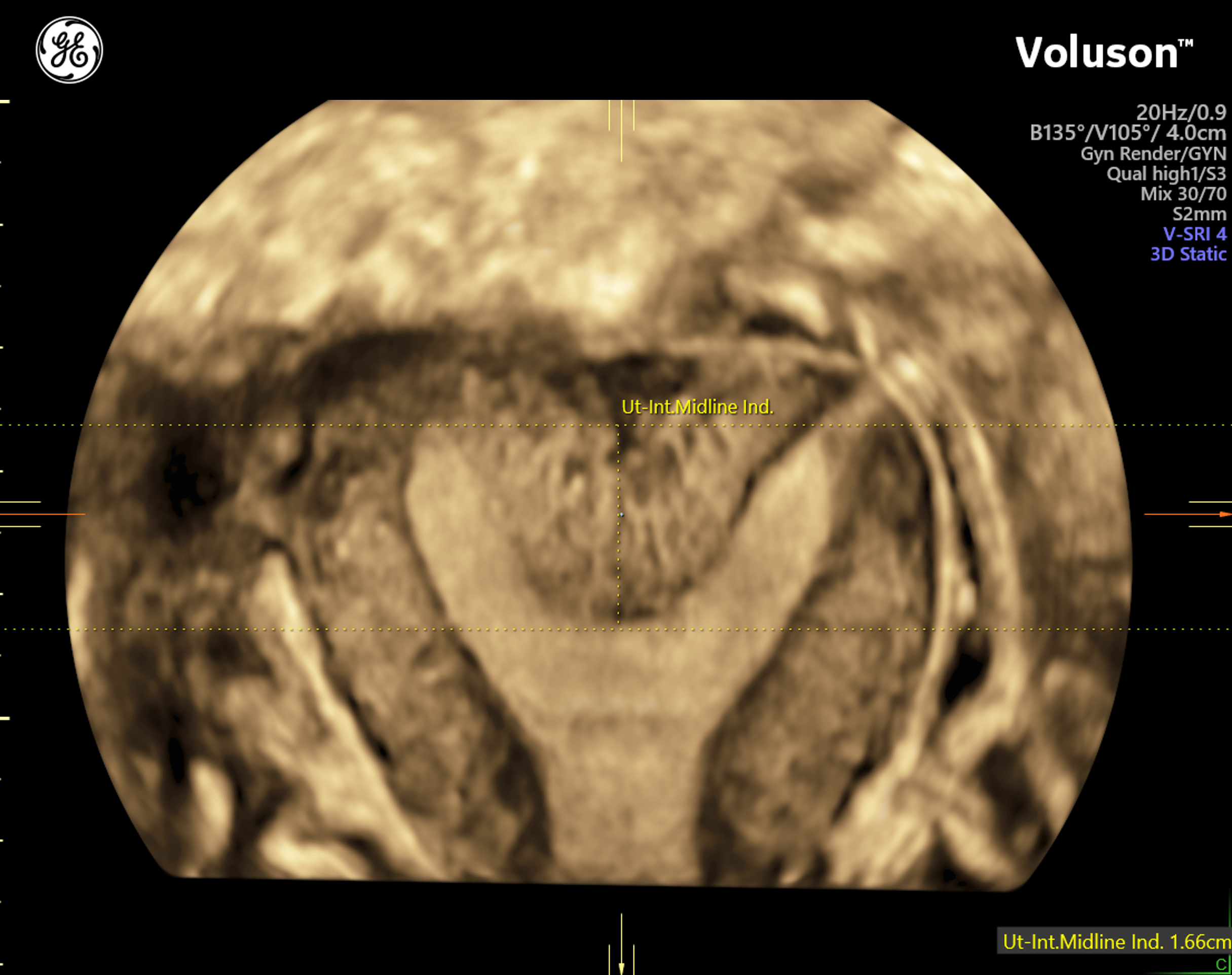
Septate uterus
Uterine fibroid and IUD, Voluson™ HDlive™ Studio technology
Volume Analysis
Once the required views are gathered, the volume contains the necessary information for analysis and correlation with other patient information. A volume may contain thousands of images for evaluation and detailed diagnostic assessment. This complete volume can be shared with a referring clinician for a second opinion or further evaluation.
The Importance of a Successful Screening
3D ultrasound enables clinicians to view planes, such as the coronal plane, not achievable with 2D imaging alone. The additional information 3D provides can help clinicians formulate a diagnosis quickly, with more confidence. 3D can aid in distinguish between conditions with similar presentations in 2D imaging. In these cases, 3D ultrasound is a useful tool for differentiating and diagnosing.
For OB/GYNs and sonographers who are learning how to accurately acquire a 3D volume, training is key. Investigate the resources your staff needs to improve their diagnostic skills by using 3D ultrasound, and research the training options offered by your ultrasound manufacturer.