Leiomyoma vs. Adenomyosis: How Ultrasound Aids in Diagnosis
When it comes to diagnosing leiomyoma vs. adenomyosis, physicians know that although these two conditions are similar in presentation, they require different treatments. Ultrasound is an integral part of gynecological assessments and can be very useful for differential diagnosis and resolving the issue.
Adenomyosis affects approximately 20 percent of reproductive age women, with a higher rate—roughly 30 to 40 percent—among those seeking treatment through assisted reproductive technology (ART). Adenomyosis has a negative effect on the outcome of in vitro fertilization, so accurate diagnosis and treatment is helpful. The condition is characterized by ingrowths of endometrial cells into the myometrium, and is typically marked by pain, menorrhagia and genitourinary symptoms.
Leiomyomas are also known as fibroids: benign smooth muscle tumors in the uterine wall that very rarely become cancer. They can cause abnormal uterine bleeding and pelvic pain. Notably, leiomyomas can co-occur with adenomyosis.
Leiomyoma vs. Adenomyosis: Key Differences
Leiomyoma and adenomyosis occur most often in women of reproductive age. Patients with one of these conditions typically present with similar symptoms: abnormal uterine bleeding, dysmenorrhea, abdominal masses, dyspareunia and infertility. 3D ultrasound is of particular help in differentiating between the two conditions.
A study in the Journal of Human Reproductive Sciences described key differences between the two disorders' appearance on transvaginal ultrasound.
For leiomyoma, two of the five following sonographic features must be present:
For adenomyosis, diagnostic criteria require two of the five following features:
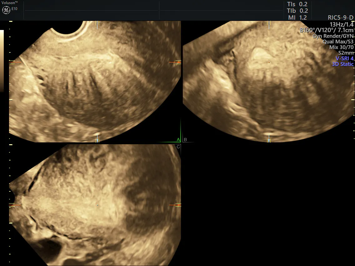
3D rendered ultrasound showing adenomyosis
Learn more about assessing the endometrium with ultrasound—download poster here.
For adenomyosis, the most common first-line treatment is conservative, including hormonal contraception or GnRH agonists depending on the patient's symptoms. For leiomyoma, or combined adenomyosis and leiomyoma, surgical options are more commonly chosen. The therapeutic interventions should depend on the patient's specific clinical challenges, such as pain, menorrhagia or infertility.
Role of Ultrasound in Differential Diagnosis
A review in SciTeMed Research on Women's Health reports that transvaginal and transabdominal ultrasound, whether alone or paired with color Doppler, are both useful in the differential diagnosis of women presenting with typical adenomyosis and leiomyoma symptoms. The study showed that 3D ultrasound had a 93.4 percent sensitivity and 95.6 percent specificity in diagnosing leiomyoma, and 95.6 percent sensitivity and 93.4 percent specificity in diagnosing adenomyosis. This means that 3D ultrasound, particularly when paired with Doppler studies, is highly valuable in the diagnosis of both conditions.
Patients who subsequently underwent treatment with surgery were included, and the radiological diagnosis was correlated with the histopathological and intraoperative diagnosis. This yielded the specificity and sensitivity of 3D ultrasound in diagnosing the conditions.
Another study, published in Fertility and Sterility, developed a clinical diagnostic prediction model to calculate the probability of adenomyosis in women suffering from dysmenorrhea and menorrhagia when transvaginal ultrasonography is used for diagnosis. The predictors included the presence of certain ultrasound features:
Each predictor is weighted based on its diagnostic significance. These criteria yielded a sensitivity of 85 percent and specificity of 78 percent in the final model.
Transvaginal ultrasonography is a key tool in distinguishing between adenomyosis and leiomyoma since both conditions present with similar symptoms. The differential diagnosis can help physicians tailor treatment to the patient's needs.
Learn more here or explore the demo today!

