

Diagnosing adenomyosis can be difficult because the vague symptoms patients often present with — chronic pelvic pain, dyspareunia and severe menstrual pain — can be linked to a number of other conditions. However, a study published by Ultrasound in Obstetrics and Gynaecology shows a strong association between the severity of pelvic pain and the number of adenomyosis features identified on ultrasound.
In order to better understand the cause of pain in patients with adenomyosis and plan their treatment, it's important to recognize the four key pain-related features of the condition that can be identified during a routine ultrasound exam.
1. Enlarged Uterus
A shift in the size of the uterus is a primary indication of adenomyosis, as long as fibroids are not present. This can present as:
- Overall uterine enlargement, where the fundas appears globular.
- An asymmetrically enlarged uterus, such as when the posterior myometrium appears larger than the anterior.
This enlargement is caused by diffuse glandular deposits within the myometrium and results in uterine tenderness, pelvic heaviness and pain.
2. Inhomogeneous Myometrial Echotexture
Adenomyosis occurs when glands and stroma from the basal layer of the endometrium migrate into the surrounding myometrium. Instead of appearing homogenous and smooth, the myometrium will appear irregular and indistinctly defined, with subendometrial lines and buds, a marked increase or decrease in echogenicity or hyperechoic islands.
These are all evidence of endometrial glands invading the myometrium and causing a hyperplastic reaction in the muscle. The stimulation of endometrial cells outside their usual location is part of what makes menstrual pain so severe for adenomyosis patients.
While some of these signs are visible on transabdominal ultrasound, a transvaginal cine sweep is especially helpful in documenting the entirety of the uterus to highlight irregularities in the myometrium.
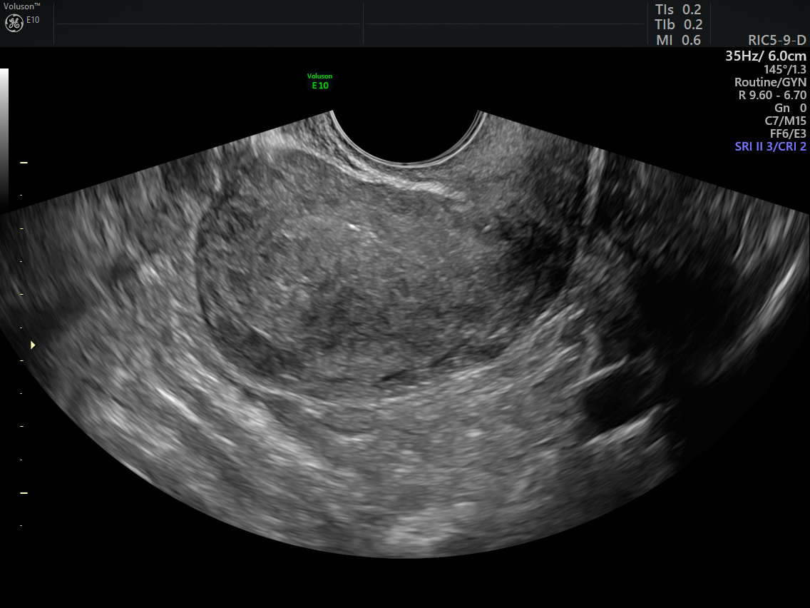
3. Cystic Anechoic Spaces in the Myometrium
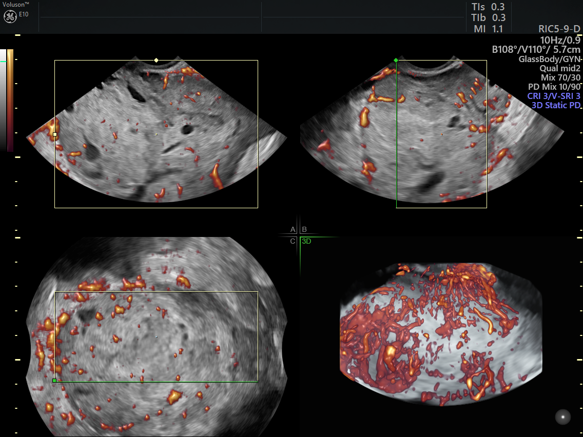
Cystic anechoic spaces are ectopic endometrial glands and stroma, which can be either distributed diffusely throughout the myometrium — diffuse adenomyosis — or as localized nodular clusters — focal adenomyosis or adenomyomas. They are variable in size and can occur throughout the myometrium. These cysts can change over time as the glands respond to hormonal levels in the body. Severe, nonresponsive dysmenorrhea is the primary symptom of myometrial cystic adenomyosis, a variation of the disease that affects mainly adolescents and young women.
Fertility and Sterility reports that transvaginal ultrasound can clearly differentiate between diffuse and focal types of adenomyosis, as well as signs of other concurrent pathology such as fibroids or endometriosis. Transvaginal ultrasound provides the clearest visualization of the myometrium, and the addition of color or power Doppler can help differentiate myometrial cysts from blood vessels.
4. Obscure Endometrial/Myometrial Border
The typically distinct junctional zone between the endometrium and myometrium can become hypoechoic, thickened and ill-defined when endometrial glands invade the surrounding tissue. 3D transvaginal ultrasound uses the acquired volume of the uterus to evaluate the junctional zone in all dimensions.
Thickness measurements have also been proposed to determine the extent of junctional zone irregularity by comparing the thickest areas with the thinnest. It's important to note that the presence of endometriosis will also affect the appearance of the junctional zone; since the two conditions may exist concurrently and cause similar symptoms, clinicians should be extra vigilant in identifying specific markers of adenomyosis.
How Distinguishing Pelvic Pain can Improve Patient Care
The positive relationship between the number of ultrasound features of adenomyosis and the degree of menstrual pain adds to the usefulness of ultrasound as an assessment tool in the search to understand severe menstrual pain. By improving their understanding of the mechanisms behind adenomyosis and pain, clinicians can help their patients find treatment as soon as possible.