

About 1 million women in the United States are affected by pelvic inflammatory disease (PID) each year. Many of these women are at risk for a serious complication called a tubo-ovarian abscess (TOA). Early detection of TOA and PID is critical to ensure the best outcomes.
Now, ultrasound is helping gynecologists uncover and evaluate how PID and TOA affect women. Learn how this technology can inform treatment and management decisions.
Exploring the Relationship of PID and TOA
Tubo-ovarian abscesses are inflammatory masses in reproductive or pelvic organs that develop as a complication of pelvic inflammatory disease. These masses can occasionally burst and lead to life-threatening sepsis.
Between 15 and 35 percent of women with PID will also develop TOA, according to Obstetrics and Gynaecology. Both conditions have a tendency to be more severe when they occur in conjunction with endometriosis.
TOA most often occurs in women of reproductive age, particularly women who have never had children. Because these abscesses are filled with bacteria, TOA can also develop secondary to another type of infection, such as appendicitis or diverticulitis.
TOA symptoms are similar to PID symptoms and may include:
- Adnexal tenderness on one or both sides
- Fever
- Elevated white blood cell count
- An active gonorrhea or chlamydia infection
- A mass that is visible on ultrasound
Tubo-Ovarian Abscess Ultrasound Detection
Ultrasound has increasingly become the preferred imaging option for evaluating and diagnosing pelvic problems. Although laparoscopy has long been considered the gold standard for diagnosing PID, ultrasound is less invasive and cheaper for patients and has a high level of sensitivity. It is also the preferred imaging tool for teen patients.
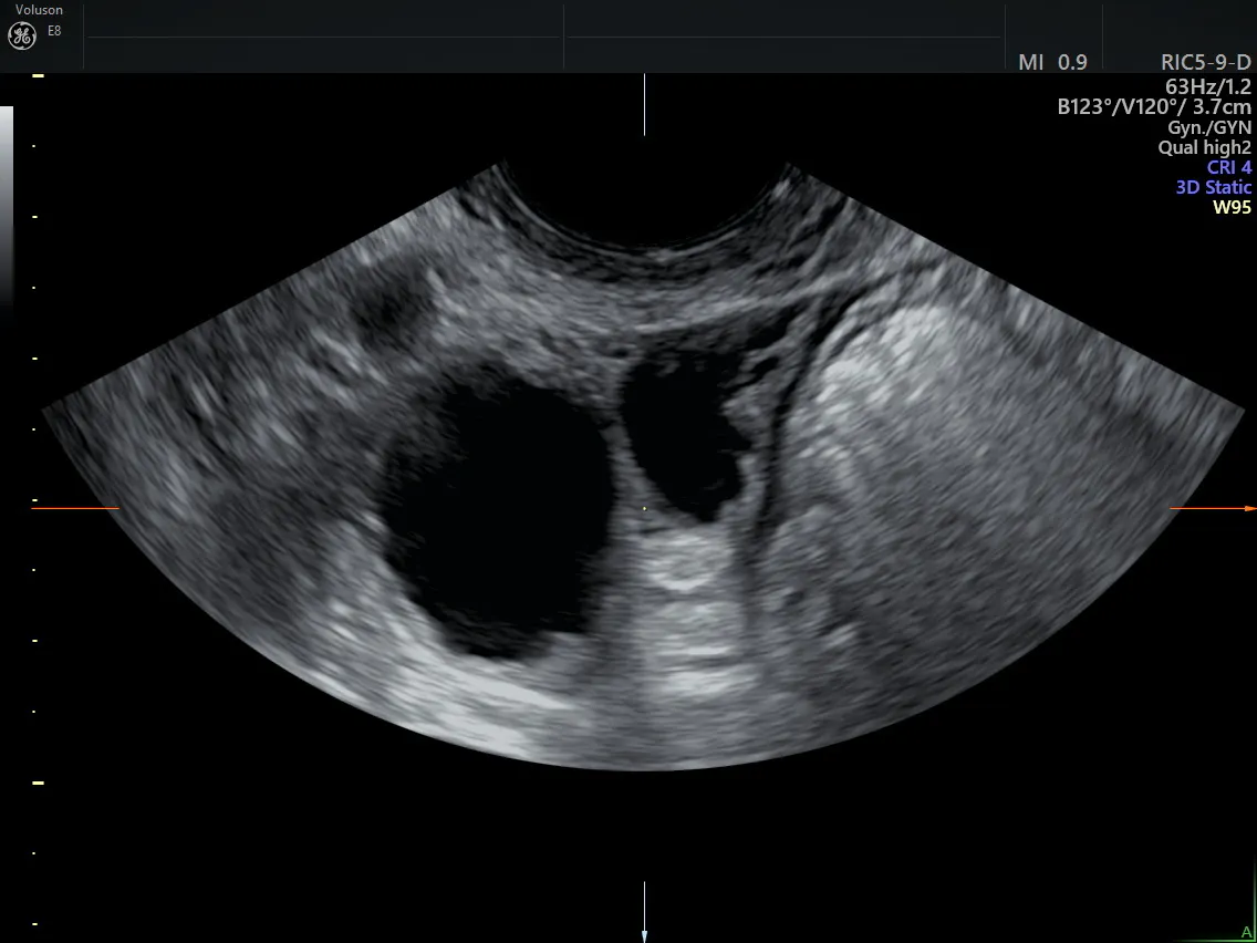
To evaluate suspected tubo-ovarian abscess symptoms, practitioners may need to perform a transabdominal or transvaginal ultrasound, depending on the location of a patient's pain. A 3D ultrasound provides a detailed view of the adnexa, the pelvis and the fallopian tubes.
TOA appears as a complex solid or cystic mass on ultrasound. Abscesses may appear on one or both sides of the ovaries. Once a clinician ascertains the size and location of any abscesses, they can use this information to inform the next steps in treatment.
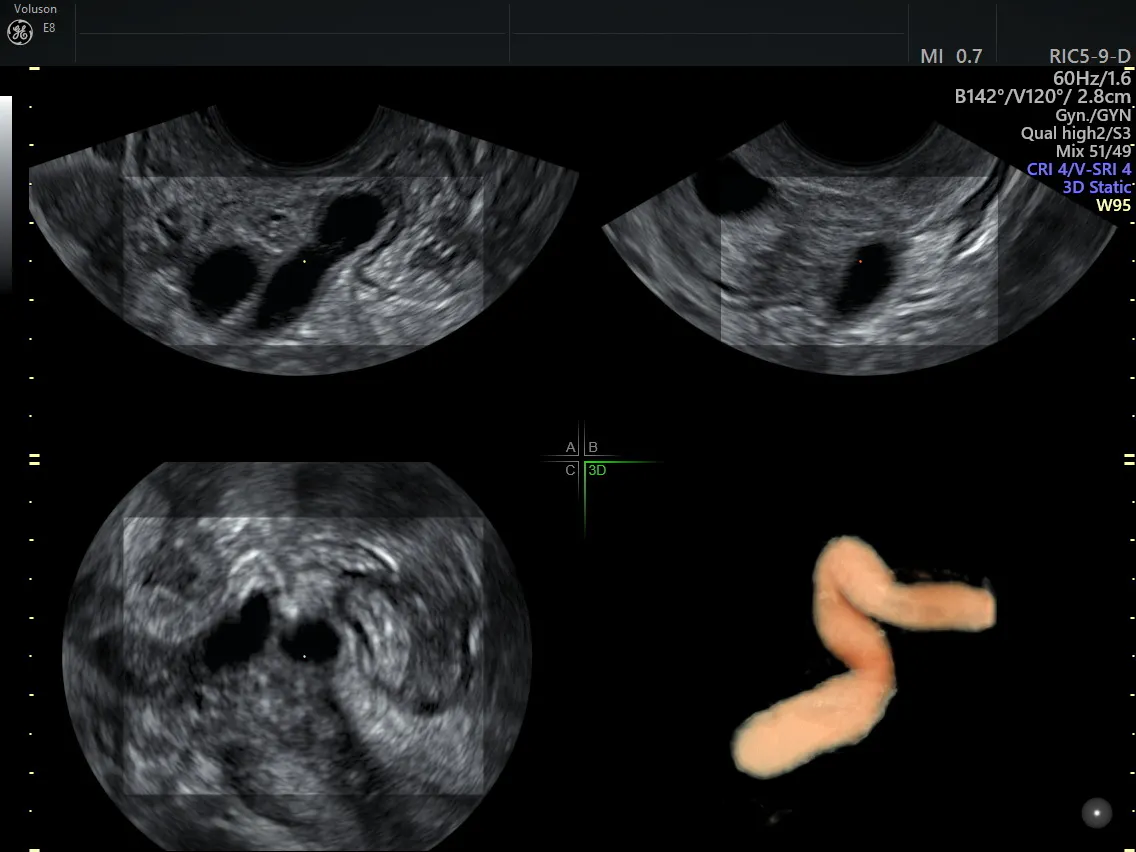
Ultrasound evaluation of hydrosalpinx
Ultrasound evaluation of hydrosalpinx
TOA Treatment and Management With Ultrasound
If sepsis is present, surgery and antibiotics should be the initial course of treatment. Antibiotics, drainage and follow-up ultrasounds may be enough to eliminate TOA if no abscesses have ruptured. However, if imaging shows that the abscesses haven't healed or the condition is worsening, further treatment will be necessary to prevent an abscess from rupturing.
Ultrasound-guided drainage of the abscess paired with antibiotics is a safe treatment plan for many women. This option also preserves fertility, which is not always possible with surgery. One study found that about 93 percent of women who underwent antibiotics and ultrasound-guided drainage successfully recovered from tubo-ovarian abscesses. Additionally, more than 60 percent of women who underwent drainage reported no pain within 48 hours.
Obstetrics and Gynaecology also reports that image-guided drainage is well-tolerated, with a shorter recovery period and less time spent in the hospital. This treatment approach also avoids the risks associated with surgery and the radiation exposure to reproductive organs that accompanies CT scans.
A growing body of research is establishing a strong role for ultrasound as a first-line diagnostic and treatment tool for PID and TOA when performed by a skilled provider or sonographer. With ultrasound, you can help patients find relief at a lower cost and with less pain and anxiety than other treatments.