Ultrasound Tutorial for Using the Uterine Trace Tool
This 3D ultrasound tutorial will help you learn how to use the Voluson Uterine Trace tool to help accurately identify uterine malformations.
Identifying uterine malformations accurately is a critical part of patient care in gynecology. Uterine anomalies, though often asymptomatic, can profoundly impact reproductive health. In one study, nearly 17% of women prone to recurrent miscarriages had congenital uterine abnormalities.
Accurate identification of uterine malformations is essential due to its direct correlation with reproductive challenges. These anomalies, which can range from septate and bicornuate uteri to arcuate deformities, can significantly impact fertility, increasing the risk of recurrent pregnancy loss and complications during gestation.
3D ultrasound is the gold standard for diagnosing uterine anomalies. According to research published in the Journal of Ultrasound in Medicine, 3D ultrasound can diagnose congenital uterine anomalies with 88% to 100% accuracy.
Getting Started with the Voluson S10 Expert Ultrasound Device
The Voluson™ Ultrasound is a 3D imaging device with advanced features and controls to enable accurate, streamlined images and results.
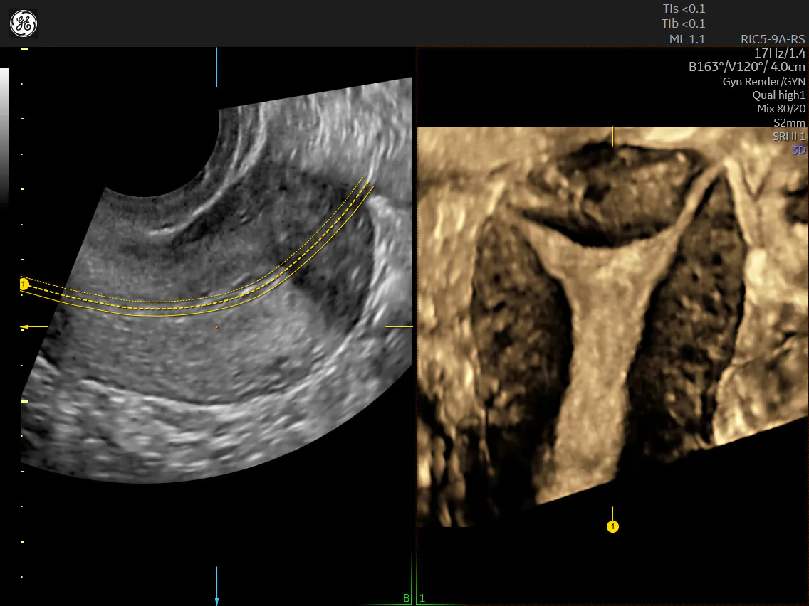
Uterine Trace is one of the tools offered on the Voluson. Incorporating 3D into your practice is easier than ever with Uterine Trace; from a standard view, simply trace the endometrium, and the system automatically acquires a volume and then exacts the critical coronal view.
Uterine Trace - OmniView VCI
Uterine Trace with Classification
How to Use the Uterine Trace Tool
To use the Uterine Trace tool of the Voluson ultrasound, follow this ultrasound tutorial:
- First, obtain a 2D image and optimize it
- Identify the mid-sagittal plane of the uterus
- Select 3D button
- Select the "Uterine Trace" preset on the touch screen
- Adjust the volume angle to include the entire uterus, up to 120°, from fundus to cervix
- Select desired quality (max=best resolution, longer acquisition time)
- Trace endometrium on touch panel, use your finger to smoothly draw a line through the cervix all the way along the endometrium to the fundus, and then release your finger
- The system will start an automated 3D sweep and will automatically acquire the volume
- The coronal view of the uterus will appear on the screen
- Confirm right/left laterality of C-Plane with 2D imaging
That's all it takes—with just three presses of a button, the ultrasound device will generate an image of the coronal plane of the uterus, the view that is most useful for identifying abnormalities.
View the full Uterine Trace demonstration using Voluson Performance HERE.
Voluson Expert - Uterine Trace - onboard education
Voluson Expert - Uterine Trace - action
Uterine Trace acquires the volume and then displays the coronal view of the uterus
Tips for Using the Uterine Trace Tool
- Include the entire uterus and cervix in the 2D sagittal image; the 3D sweep starts automatically when endometrium trace is finished
- Watch as volume is being acquired to be assured entire uterus is included in the volume; if entire uterus is not included, the Volume Angle may need to be adjusted
- Adjust position and rotation of line as needed
- VCI (Volume Contrast Imaging) is added according to selected preset for additional contrast resolution; the thickness may be adjusted
- To ensure complete visualization of cornua bilaterally, consider aligning endometrium in B-Plane in Multiplanar view
- Select "Uterine Classification"
- Select uterine cavity icon based on ESHRE or ASRM classification guidelines
Interpreting the Results
The resulting coronal view that the Uterine Trace tool displays is key to diagnosing uterine abnormalities. This is the plane in which both internal (endometrial) and external (serosal surface) contours of the uterine fundus can be visualized simultaneously, thus facilitating the diagnosis.
For example, septate uteri, characterized by a septum dividing the uterine cavity, can be readily visualized in the coronal plane. The depth and extent of the septum can be measured accurately as well. Similarly, bicornuate uteri, where the uterus is divided into two separate horns, are clearly seen in the coronal view. This view facilitates the measurement of the angle and degree of separation between the horns, which can help predict potential health outcomes for patients during pregnancy.
Uterine Classification
Uterine Trace - Septate Uterus
Likewise, the coronal view provides essential insights into unicornuate uteri, where only half the uterus develops, and didelphic uteri, where it is divided into two completely separate structures. The length and configuration of these irregularly shaped structures can be measured accurately with the 3D coronal view that is generated by the Uterine Trace tool.
The Uterine Trace tool integrates with ASRM Uterine Classification guidelines as well as the ESHRE/ESGE Uterine Classification guidelines. These classification systems have defined measurement techniques, reference points, and specific cut-offs to differentiate between normal uteri and those with abnormalities.
The use of 3D ultrasound, and specifically the coronal view, has significantly enhanced gynecology's ability to easily and accurately diagnose congenital uterine abnormalities. Hopefully this tutorial has informed your use of the Uterine Trace tool and assisted in use of 3D ultrasound imaging in gynecology. As medical imaging continues to evolve, the integration of 3D ultrasound into women's health care promises to further refine medicine's ability to diagnose and manage uterine abnormalities, ultimately enhancing patient outcomes.

