
Overactive bladder symptoms and other forms of voiding dysfunction affect up to 43 percent of women and are often associated with underlying disorders such as pelvic organ prolapse, according to the Canadian Urological Association Journal. Overactive bladder dysfunction can cause urgency, frequency, nocturia and incontinence.
Many cases yield symptoms that lead to embarrassment and disrupt work schedules, social interactions and other everyday activities. This condition can be severe enough to cause depression. Therefore, it is important to find noninvasive solutions to these conditions in urogynecology to help patients manage their symptoms.
Using Real-Time Ultrasounds in Urogynecology
Physicians can only treat patients with overactive bladders after they have gained a better understanding of how the bladders fill and empty. Real-time bladder volume ultrasound results can provide urogynecologists with accurate and precise serial measurements of bladder volumes during filling and of residuals after bladder emptying.
Post-void residual volume (PVR) is commonly used to diagnose urinary retention, detrusor underactivity and other bladder impairments. PVR measurement is an essential aspect of all urogynecology assessments, particularly when diagnosing patients who experience voiding difficulty — abnormally slow or incomplete voiding, for example — prior to surgical intervention for urodynamic stress incontinence.
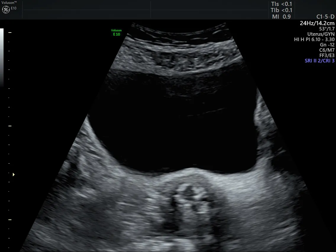
Abnormal bladder ultrasound image - urinary bladder
Choosing 3D Ultrasound to Assist in Urogynecology Assessment
An abnormal bladder ultrasound gives clinicians a noninvasive diagnostic method that is both quick and accurate, especially with the development of 3D ultrasound volume-measuring software. The International Continence Society has reported that a 3D ultrasound measures a wide range of bladder volume in a reproducible and accurate manner, making it superior to 2D ultrasound.
However, physicians can also estimate bladder volume accurately with 2D ultrasound using transabdominal, transvaginal or translabial approaches. Ultrasound in Obstetrics and Gynecology reports that in cases of women with advanced pelvic organ prolapse, translabial 2D ultrasound most accurately measures bladder volume in both pre- and post-void states.
Managing Overactive Bladder Syndrome
Estimating pre- and post-void residual urine volume is useful when diagnosing and managing patients who suffer from an overactive bladder and voiding dysfunction. This tool provides a noninvasive option for physicians looking to avoid the trauma, discomfort and potential for infection associated with measuring bladder volume by urethral catheterization.
Ultrasonographic measurements of bladder volume can offer significant ease and reproducibility of measurement in all ranges of possible bladder volume. Both 3D volume and 2D conventional ultrasound can help physicians better estimate selected bladder volumes to assist with presurgical planning, pelvic organ prolapse and overactive bladder syndrome.


