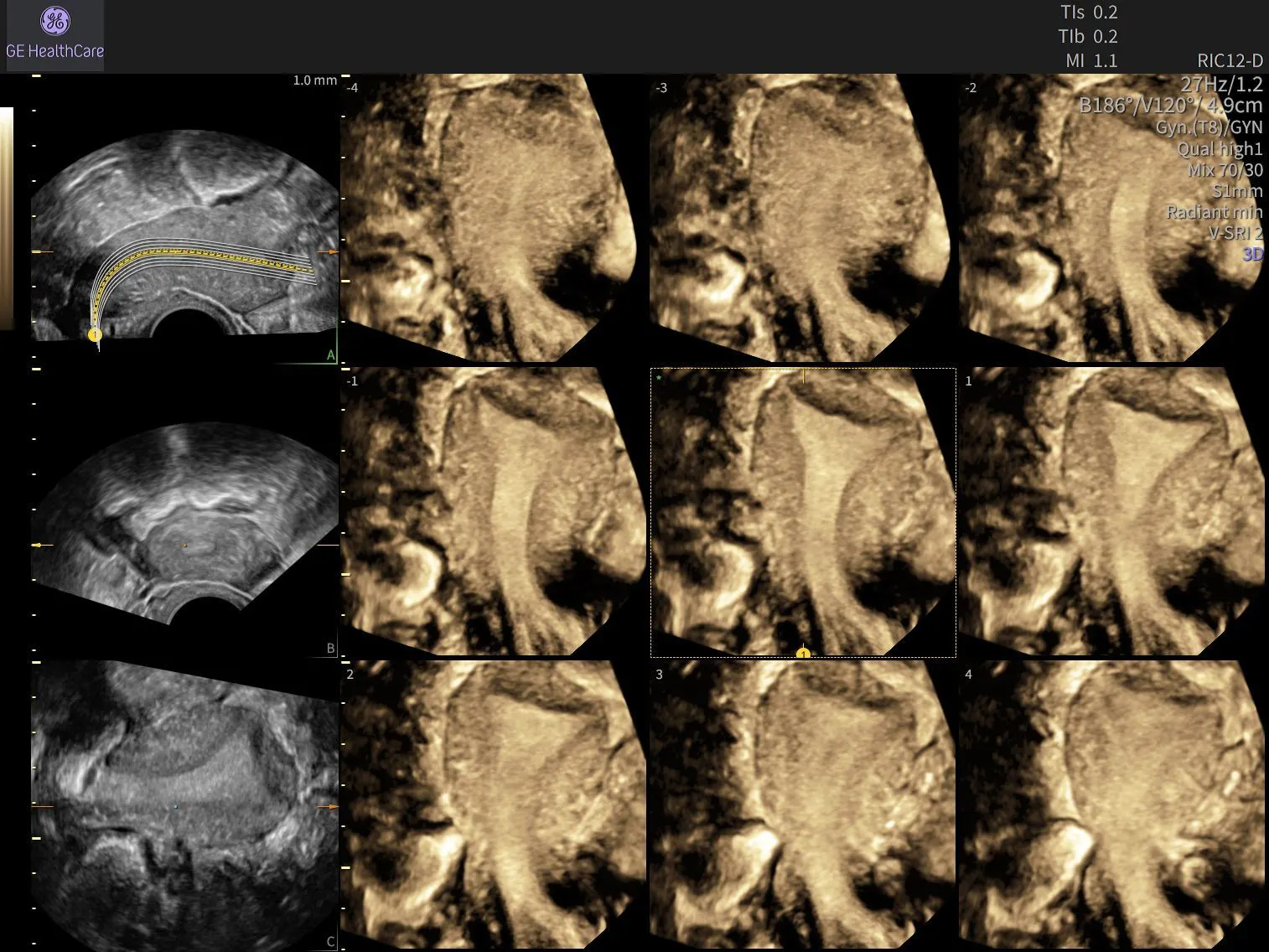
Earlier Insights in Early Pregnancy: Supporting the Assessment of Pregnancy of Unknown Location and Ectopic Pregnancy Risk
Pregnancy of unknown location (PUL) is a clinical finding that occurs when a pregnancy test is positive, but no intrauterine or extrauterine pregnancy can be visualised on transvaginal ultrasound. Most PUL cases ultimately resolve as either a viable intrauterine pregnancy or a miscarriage without requiring intervention.
However, a significant minority of cases require closer evaluation. Extrauterine pregnancies may be present in up to 30% of PUL cases, with ectopic pregnancy occurring in approximately 8% to 16%.¹˒² Accurately identifying patients who require follow-up is essential for supporting early diagnosis, avoiding unnecessary intervention, and helping improve maternal outcomes.
By combining ultrasound assessment with evidence-based risk stratification tools, clinicians can gain valuable insights that support informed decision-making during a critical stage of early pregnancy care.
Understanding Pregnancy of Unknown Location
Patients with a PUL often present in early pregnancy with symptoms such as pelvic pain, vaginal bleeding, or both, alongside a confirmed pregnancy based on serum human chorionic gonadotropin (hCG) levels. Despite these findings, transvaginal ultrasound demonstrates neither an intrauterine gestational sac nor evidence of an extrauterine pregnancy.
At this stage, determining the location and viability of the pregnancy can be challenging. Clinicians rely on a combination of laboratory testing and serial ultrasound assessment to evaluate the risk of ectopic pregnancy and guide further management.¹
Early detection remains particularly important because ectopic pregnancy continues to be one of the leading causes of maternal mortality in early pregnancy. Although ultrasound plays a central role in evaluation, ectopic pregnancies can be difficult to visualise at an early stage. Risk stratification therefore depends on integrating ultrasound findings with biochemical markers to guide appropriate follow-up and intervention.
Looking Beyond a Single hCG Measurement
Historically, clinicians used the hCG discriminatory zone as a tool for determining the likelihood of visualising an intrauterine pregnancy on ultrasound. However, reliance on a single hCG measurement is no longer recommended because it may result in inappropriate intervention, including administration of methotrexate in potentially viable pregnancies.³
The discriminatory zone is based on the assumption that when hCG levels reach approximately 1,000 to 1,500 IU/L, an intrauterine pregnancy should be visible on transvaginal ultrasound. If no pregnancy is identified, concern for an extrauterine pregnancy increases. However, clinicians with advanced ultrasound expertise may sometimes identify ectopic pregnancies at hCG levels around 1,000 IU/L.³
Rather than relying on a single result, serial measurements provide greater clinical value. The hCG ratio compares serum hCG concentrations obtained 48 hours apart and can help support risk assessment.
- An hCG ratio of less than 0.87 is generally associated with a failing PUL that is unlikely to require intervention.
- An hCG ratio greater than 1.66 is suggestive of a viable intrauterine pregnancy.
- Ectopic pregnancy remains possible, however, even when the hCG ratio is 2.0 or greater.⁴
Importantly, no single hCG pattern reliably identifies ectopic pregnancy. Many ectopic pregnancies demonstrate rising hCG levels, highlighting the importance of combining biochemical trends with ultrasound findings and clinical evaluation.⁴
Advancing Risk Stratification
Risk prediction models are increasingly helping clinicians assess patients with PUL more effectively.
The M4 model incorporates the initial serum hCG concentration and the hCG ratio to estimate the likelihood of ectopic pregnancy.⁵
The more recent M6 model expands upon this approach by incorporating serum progesterone levels alongside initial hCG measurements and hCG ratio. This allows clinicians to generate probabilities for:
- Viable intrauterine pregnancy
- Failed PUL
- Ectopic pregnancy⁵
Both models have demonstrated high sensitivity and strong negative predictive value for identifying women who may be suitable for expectant management and ongoing observation.⁴˒⁵
When used alongside ultrasound assessment, these tools can support more targeted follow-up pathways and help identify patients who may benefit from additional imaging to establish pregnancy location.
The Critical Role of Ultrasound in Ectopic Pregnancy Assessment
Ultrasound remains central to the evaluation and monitoring of patients at risk of ectopic pregnancy.
Women with suspected ectopic pregnancy can be triaged into follow-up programmes that include serial ultrasound examinations. In some cases, ultrasound may demonstrate a gestational sac, embryo, or even fetal cardiac activity located outside the uterine cavity, although these findings are present in only a minority of cases. More commonly, clinicians may identify free pelvic fluid or a non-homogeneous adnexal mass.⁴
A classic tubal ectopic pregnancy often appears as an adnexal mass separate from the ovary. Recognition of these findings can help support earlier diagnosis and intervention before rupture occurs.
Cervical ectopic pregnancy presents unique sonographic features, including:
- An empty uterine cavity
- A barrel-shaped cervix
- A gestational sac located within the cervix below the internal cervical os
- Absence of the sliding sign
- Peritrophoblastic blood flow surrounding the gestational sac on colour Doppler imaging⁶
By combining clinical assessment, biochemical monitoring, and high-quality ultrasound evaluation, clinicians can improve diagnostic confidence and support timely management decisions.
Recognising Pitfalls and Atypical Presentations
Not all PUL cases follow a typical presentation.
Heterotopic pregnancy presents a particular diagnostic challenge. Although rare in spontaneous conception, it occurs considerably more frequently following assisted reproductive technologies, with reported incidences approaching 1 in 100 pregnancies in this population.⁷
A key consideration is that identification of an intrauterine pregnancy does not exclude the presence of a concurrent ectopic pregnancy. Careful ultrasound evaluation therefore remains essential even when an intrauterine gestation has been confirmed.
Interstitial (cornual) ectopic pregnancies also require particular attention. Because these pregnancies are surrounded by myometrium, they may be more difficult to recognise and often present later than ampullary ectopic pregnancies. This delayed presentation can increase the risk of haemorrhage and associated complications.⁸
Another common diagnostic pitfall is the pseudogestational sac. This fluid collection within the uterine cavity may resemble an early gestational sac but lacks the distinguishing features of a true intrauterine pregnancy and may occur in association with ectopic pregnancy.⁹
Careful transvaginal ultrasound assessment can help differentiate these findings. One particularly valuable ultrasound marker is the double decidual sac sign (DDSS), visualised as two concentric echogenic rings surrounding an endometrial fluid collection. The presence of the DDSS supports the diagnosis of an intrauterine pregnancy, whereas only a single ring may be seen in ectopic pregnancy.¹⁰
Bringing Together Risk Stratification and Ultrasound Insights
Currently, no international consensus exists regarding the optimal follow-up strategy for all women with a pregnancy of unknown location.
Management should be individualised according to clinical presentation, risk profile, biochemical trends, and ultrasound findings. Women assessed as low risk may be appropriate candidates for expectant management, while those at higher risk of ectopic pregnancy often require additional hCG monitoring and serial ultrasound examinations.
Intervention should be considered when:
- hCG levels plateau or rise abnormally
- Ultrasound findings become suspicious for ectopic pregnancy
- Clinical symptoms worsen
- Haemodynamic instability develops
- Signs of rupture are present⁶˒¹¹
Expectant management remains a safe option for appropriately selected women with resolving ectopic pregnancy. Medical or surgical treatment is indicated when spontaneous resolution is unlikely or when clinical risk increases.⁶˒¹¹
Empowering Earlier Decisions in Early Pregnancy Care
Every ultrasound examination provides an opportunity to generate valuable clinical information.
For women presenting with a pregnancy of unknown location, combining transvaginal ultrasound with evidence-based risk stratification models can support earlier identification of ectopic pregnancy while helping reduce unnecessary interventions.
As ultrasound technology, clinical guidelines, and practitioner expertise continue to evolve, clinicians are increasingly equipped to evaluate complex early pregnancy presentations with greater confidence.
Through ongoing education, systematic scanning approaches, and thoughtful integration of clinical and imaging data, healthcare professionals can support earlier recognition of ectopic pregnancy, more informed management decisions, and improved maternal outcomes.
References
1.Kovacs P. Pregnancy of Unknown Location. Medscape. Published February 5, 2019. Accessed July 1, 2026. https://www.medscape.com/viewarticle/908491?form=fpf
2.Van Calster B, Abdallah Y, Guha S, et al. Rationalizing the management of pregnancies of unknown location: temporal and external validation of a risk prediction model on 1962 pregnancies. Hum Reprod. 2013;28(3):609-616. doi:10.1093/humrep/des440
3.Bobdiwala S, Al-Memar M, Farren J, Bourne T. Factors to consider in pregnancy of unknown location. Womens Health (Lond). 2017;13(2):27-33. doi:10.1177/1745505717709677
4.Boyraz G, Bozdağ G. Pregnancy of unknown location. J Turk Ger Gynecol Assoc. 2013;14(2):104-108. Published 2013 Jun 1. doi:10.5152/jtgga.2013.74317
5.Bobdiwala S, Christodoulou E, Farren J, et al. Triaging women with pregnancy of unknown location using two-step protocol including M6 model: clinical implementation study. Ultrasound Obstet Gynecol. 2020;55(1):105-114. doi:10.1002/uog.20420
6.Diagnosis and Management of Ectopic Pregnancy: Green-top Guideline No. 21. BJOG. 2016;123(13):e15-e55. doi:10.1111/1471-0528.14189
7.Maleki A, Khalid N, Rajesh Patel C, El-Mahdi E. The rising incidence of heterotopic pregnancy: Current perspectives and associations with in-vitro fertilization. Eur J Obstet Gynecol Reprod Biol. 2021;266:138-144. doi:10.1016/j.ejogrb.2021.09.031
8.Sharma C, Patel H. Ruptured Cornual Ectopic Pregnancy: A Rare and Challenging Obstetric Emergency. Cureus. 2023;15(10):e47842.doi:10.7759/cureus.47842
9.Bunjamin IA, Gianina K, Nisa AS, et al. Diagnostic Pitfall: Intrauterine Pseudogestational Sac Mimicking an Anembryonic Pregnancy in Tubal Ectopic Pregnancy A Case Report. Int J Womens Health. 2026;18:582019. Published 2026 Mar 7. doi:10.2147/IJWH.S582019
10.Richardson A, Hopkisson J, Campbell B, Raine-Fenning N. Use of double decidual sac sign to confirm intrauterine pregnancy location prior to sonographic visualization of embryonic contents. Ultrasound Obstet Gynecol. 2017;49: 643-648. doi: 10.1002/uog.15966
11.Mol F, Mol BW, Ankum WM, van der Veen F, Hajenius PJ. Current evidence on surgery, systemic methotrexate and expectant management in the treatment of tubal ectopic pregnancy: a systematic review and meta-analysis. Hum Reprod Update. 2008;14(4):309–319. doi:10.1093/humupd/dmn012

