
The use of assisted reproductive technologies (ARTs), specifically in vitro fertilization (IVF), can be a primary risk factor for ectopic pregnancy (EP). According to one studyin Reproductive Biology and Endocrinology, ectopic pregnancy occurs in some 2 to 5 percent of all IVF pregnancies, as opposed to less than 2 percent of those achieved without fertility treatment. Unlike some other pregnancy complications, the signs of an ectopic pregnancy — such as nausea and breast tenderness — are often either absent or written off as normal pregnancy symptoms.
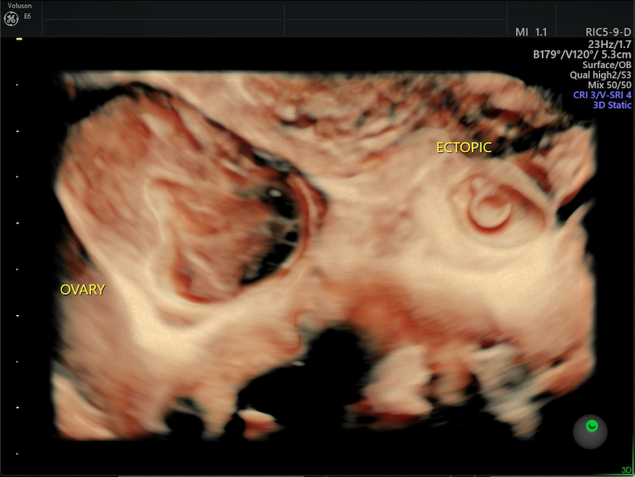
Ectopic pregnancy adjacent to the ovary.
Ectopic pregnancy adjacent to the ovary.
However, with new advances in ultrasound (US) technology, doctors now have the chance to more closely monitor the position of embryos earlier in assisted pregnancies, potentially making the use of IVF much safer for patients and decreasing their ectopic pregnancy risk. Most physicians are already familiar with 2D ultrasound technology, which creates images of internal structures or cavities using high-frequency sound waves. However, 2D ultrasound only presents images from the sagittal and transverse planes of the body. In contrast, 3D and 4D ultrasound also capture images from the coronal plane, giving physicians a more complete view of the internal organs.
Better Visualization with 3D/4D Ultrasound
Embryo transfer is the cornerstone of ART. Before the procedure, a predetermined number of embryos are loaded into a fine transfer catheter that is then inserted into a patient's uterus through the vagina and cervix. In your own practice, you may use transvaginal or transabdominal ultrasound to guide placement of the catheter tip inside the uterus.
In recent years, studies have suggested that embryo placement in the lower or central part of the uterus has a greater likelihood of achieving pregnancy. While ultrasound devices can certainly help physicians visualize the catheter tip in the uterus, some research has shown discrepancies in the perceived catheter tip location and the actual position of the catheter tip when viewed using 2D US as opposed to 4D or 3D ultrasound.
Additionally, it's now thought that visualizing air bubblesthat are implanted along with the embryos, referred to as the embryo flash, is more predictive of a successful pregnancy than the placement of the catheter tip inside the uterus. These air bubbles are easily visualized along the coronal plane using 3D ultrasound, a feature 2D systems cannot replicate.
Diagnosing Ectopic Pregnancy With Ultrasound
Even with better visualization techniques, the possibility of embryo migration within the uterus after a transfer still exists. It is possible for embryos to move a significant distance away from the original transfer site, sometimes within a short period of time. In some cases, this migration leads to an ectopic pregnancy. Be transparent with your patients about this rare but possible complication.
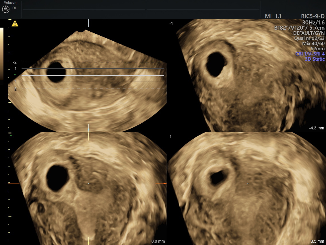
Tomographic Ultrasound Imaging (TUI) of uterine coronal plane with ectopic pregnancy
Tomographic Ultrasound Imaging (TUI) of uterine coronal plane with ectopic pregnancy.
Since 3D ultrasound allows physicians to view structures in the coronal plane, this imaging technique provides a complete view of the anatomy of the reproductive organs. You can easily view structures surrounding the uterus, such as the fallopian tubes, for evidence of any abnormalities. Additionally, 3D and 4D ultrasound can be used to visualize the embryo flash over a period of time, suggesting a migratory pattern you can use to predict the site of implantation.
If the evidence suggests migration into a fallopian tube, you can easily confirm your diagnosis using advanced ultrasound technologies. One study found that the sensitivity and specificity of an initial ultrasound to detect ectopic pregnancy were 88.5 and 96.5 percent respectively.
Ectopic pregnancy risk is a real concern for you and your patients, but advances in ultrasound technology can help you recognize and diagnose this potentially life-threatening complication sooner. Better ultrasound technologies may also help you transfer embryos more easily to a suitable location in the uterus. And once a transfer is complete, 3D and 4D ultrasound technology allows you to more accurately monitor the embryo's progression to implantation than ever before.


