

Uterine malformations are reproductive malformations that stem from abnormal growth during fetal development. Also known as congenital uterine anomalies and Müllerian duct anomalies, these malformations can increase the risk of infertility or adverse pregnancy outcomes, such as miscarriage, intrauterine growth restrictions and preterm delivery.
Tools such as 3D ultrasound can help clinicians identify and evaluate a uterine malformation and determine the best treatment options.
Understanding Each Type of Uterine Malformation
The European Society of Human Reproduction and Embryology (ESHRE) and the European Society for Gynaecological Endoscopy (ESGE) group malformations into several different categories.
The most common form of congenital uterine anomaly is a septate uterus, which occurs when the Müllerian ducts fail to completely fuse during development. There are two types of septate uteruses:
- Complete septate uterus, in which the septum divides the whole uterine cavity.
- Subseptate uterus, a partial separation of the uterine cavity ending above the cervix.
A study in the journal Human Reproduction Update found that about 8 percent of infertile women have a septated uterus, as do 13 percent of those with a history of miscarriage.
Other types of uterine malformations include unicornuate (one-horned) uterus and bicornuate (two-horned) uterus. The prevalence of pregnancy risk and reduced fertility varies with the type of malformation. Overall, the presence of a congenital uterine malformation is associated with a 15 percent lower probability of conception, according to Female Genital Tract Congenital Malformations.
3D Ultrasound for Uterine Malformations
Although ultrasound has been used to help evaluate uterine malformations for decades, its success used to be variable. More invasive and less accurate approaches, such as hysterosalpingography (HSG) and laparoscopy, were once necessary to establish a diagnosis.
HSG alone is only 55 percent effective at differentiating a bicornuate vs. septate uterus. It is also painful for patients and carries the risks of contributing to pelvic inflammatory disease (PID) and exposing patients to contrast materials, according to the International Journal of Fertility and Sterility.
Fortunately, the advent of 3D ultrasound technology has dramatically improved the diagnosis and treatment of uterine anomalies. This approach allows clinicians to view the coronal plane, or the "slice" of the body between the back and front, and create 3D ultrasound images that more realistically represent the internal organs.
3D ultrasound offers a number of advantages over traditional diagnostic methods. An increasing body of evidence shows that this technology accurately identifies uterine malformations — more so than 2D ultrasound, magnetic resonance imaging and other traditional evaluation methods. What's more, 3D ultrasound is minimally invasive and lacks many of the risks and side effects associated with HSG and laparoscopy.
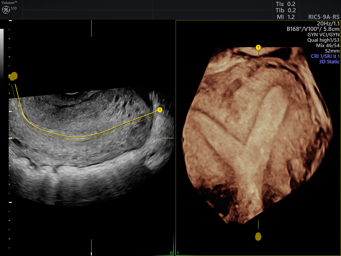
Ultrasound of subseptate uterine malformation using OmniView technology
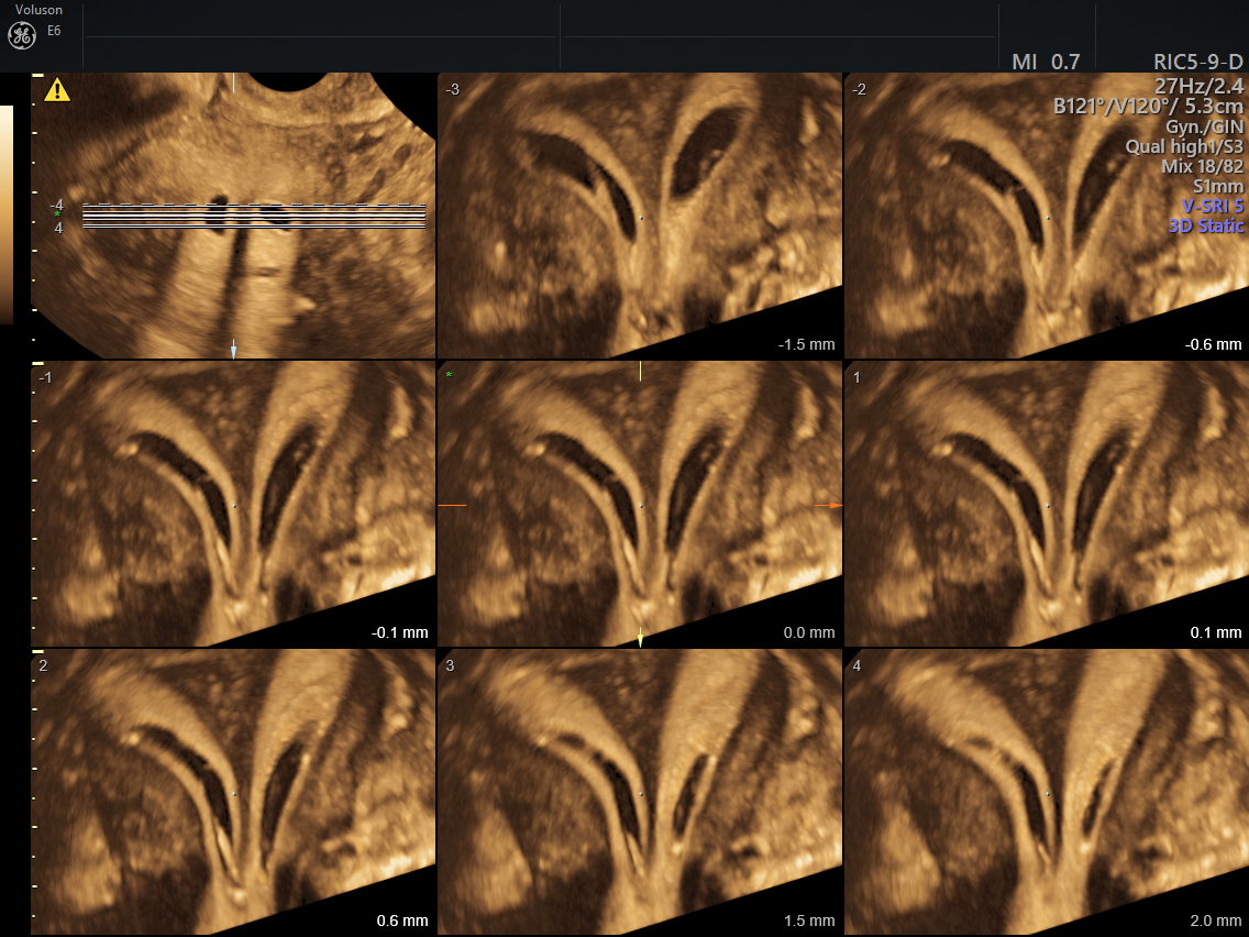
Sonohysterogram of septate uterine malformation using TUI
Using 3D Technology Beyond Diagnosis
Along with its role in accurately identifying and classifying uterine malformations, 3D ultrasound can also aid treatment.
Options for boosting fertility in women with congenital uterine anomalies include laparotomy and hysteroscopic techniques. In recent years, 3D ultrasound has been increasingly used during hysteroscopic surgery to confirm uterine contour, decrease the risk of uterine perforation, and assess the complete removal of the septum and the presence of other uterine anomalies.
The pregnancy-related risks of these malformations, such as reduced fertility and a higher chance of miscarriage, can be extremely stressful for women. Gynecologists can play an important role in caring for and supporting patients through the diagnosis and treatment process. With the use of 3D ultrasound, the evaluation, diagnosis and even treatment of uterine anomalies can become a simpler and more comfortable experience for patients.


