

Urogynecology focuses on understanding, diagnosing and treating problems with the female pelvic floor, most notably handling issues involving pelvic organ prolapse, incontinence and bowel or bladder dysfunction. Ultrasound systems have been a growing tool in the field and their potential uses have greatly increased in the past two decades. However, they are often underused in urogynecology, especially in the United States.
The development of the urogynecology ultrasound has expanded physicians' and researchers' understanding of pelvic floor disorders and has become their top choice for diagnostic imaging. Ultrasounds also offer a cost-effective choice for patients looking for a noninvasive diagnostic tool.
Evolution of Ultrasound in Urogynecology
Back in 1999, a review published in the International Urogynecology Journal recommended that physicians use ultrasounds to examine the upper and lower urinary tract. The low cost of ultrasound installation and performance relative to other imaging modalities it is recommended as a screening procedure in urogynecology. The authors looked forward to further development of 3D ultrasounds and transvaginal color Doppler to gain more understanding of women's pelvic floor issues, as well as increased use in the office for screening.
Jumping ahead nearly 10 years, a similar review in the Journal of Medical Ultrasound highlighted advances in pelvic floor imaging and further uses of ultrasound technology. The authors advocated for standardized guidelines for urogynecology ultrasound systems.
With continued research, adoption of ultrasounds to evaluate pelvic floor dysfunction has grown. The National German Guideline now cites pelvic floor ultrasounds as the "gold standard in gynecology for the morphological diagnosis of incontinence and of functional disorders of the pelvic floor."
Growing Uses for Urogynecology Ultrasound
As technology has advanced, researchers and physicians have found more ways to use ultrasounds. At first, they were primarily used to evaluate the bladder and bladder neck. Uses then expanded to examining prolapse as well as mesh and slings. Current research is looking toward evaluating pelvic floor dysfunction after childbirth and the role of 4D ultrasound. Currently, physicians can utilize urogynecology ultrasound technology to:
- Assess upper urinary tract lesions, bladder neck mobility and urethra.
- Measure post-void residue.
- Diagnose vaginal cysts, urethral diverticula and rectal intussusception.
- Visualize pelvic organ prolapse defects.
- Visualize and evaluate mesh implants and slings.
- Measure bladder wall thickness.
- Preoperative and post-operative imaging.
- Evaluate anal incontinence and anal and rectal tumors.
- Evaluate pelvic floor dysfunction after childbirth.
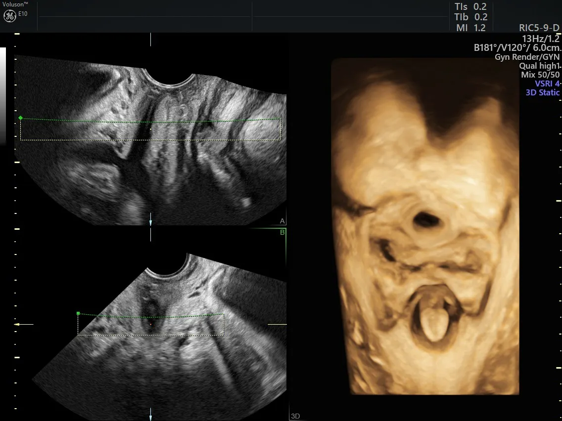
3D Ultrasound - Pelvic Floor
2D vs. 3D vs. 4D Ultrasound
Urogynecology ultrasound systems do require a degree of knowledge and training to perform the different types of exams. The ultrasound may be done as transabdominal, introital, transperineal, endo-anal, transvaginal, transrectal or translabial. In some cases, such as urinary incontinence, a 2D ultrasound may be sufficient to visualize the pelvis. In other cases, such as visualizing the pelvic floor muscles after childbirth, 3D ultrasound is necessary to view the planes of the muscles. Applications for 4D ultrasound are still being researched for their uses in practice, according to a review in Obstetrics and Gynecology.
A growing body of research supports the continued use of ultrasound in urogynecology. It provides a simple, inexpensive way to view, evaluate and diagnose pelvic floor dysfunction and is performing on par with traditional imaging modalities such as X-ray and MRI. Increased use is leading to more standardization, training and guidelines around the world.