
Nearly 5 percent of women in the United States live with pelvic inflammatory disease (PID), according to the U.S. Department of Health and Human Services. The condition is most common in people age 15 to 24, although any sexually active woman can develop PID. For pelvic inflammatory disease ultrasound may be the key to an accurate diagnosis when a patient presents with suspicious symptoms.
Generally, pelvic inflammatory disease results from an ascending lower genital tract infection, such as many sexually transmitted infections. However, other issues, such as adnexal masses, may contribute to this condition. Other than age, major risk factors include:
- Intrauterine devices.
- Multiple sexual partners.
- Prior uterine procedures.
Signs of PID
Women with suspected PID often present with a variety of symptoms, including:
- Pelvic pain.
- Fever.
- Heavy vaginal discharge.
- Dyspareunia.
- Difficulty becoming pregnant.
- Leukocytosis.
However, other vague symptoms such as fatigue or bowel discomfort often occur along with traditional signs of the condition. Clinical examination should include a complete pelvic exam, together with laboratory work and imaging.
Traditionally, CT scans are used to assess the extent of any peritoneal disease. Gynecologists may choose to obtain CT scans of a patient if suspected PID is recurrent or chronic in nature. Additionally, magnetic resonance imaging (MRI) may prove useful in evaluating the uterine and adnexal structures as part of a differential diagnosis.
Pelvic Inflammatory Disease Ultrasound Makers
Ultrasound has emerged as a useful imaging modality for the evaluation of pelvic inflammatory disease because it is easily accessible and generally cheaper than other imaging choices. Most gynecological offices already have ultrasound equipment available, making same-day examination possible.
Though transabdominal ultrasound is sometimes a viable option, transvaginal ultrasound is preferable for evaluating patients with suspected PID for any adnexal masses, which sometimes accompany the condition. According to a study in Case Reports in Obstetrics and Gynecology, transvaginal ultrasound shows a 93 percent sensitivity and an 81 percent specificity when using the International Ovarian Tumour Analysis "Simple Rules" classification system.
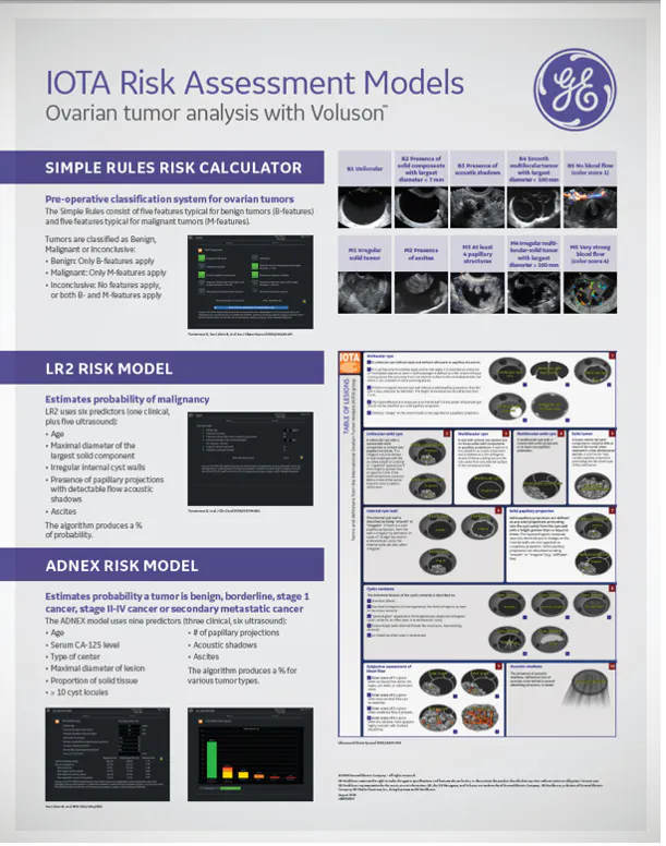
Iota Risk Models
IOTA risk models for ovarian tumor analysis
Aside from adnexal masses, transvaginal ultrasound can reveal several distinctive findings that may indicate PID, including:
- Heterogenous (tubo ovarian abscess can have solid and cystic components), low-level echogenic areas in the adnexal regions or ovaries .
- Free fluid in the Pouch of Douglas.
- Hydrosalpinx.
- A "sliding sign" indicating obliteration of the Pouch of Douglas.
- "String of Pearls" or Cogwheel.
<strong>
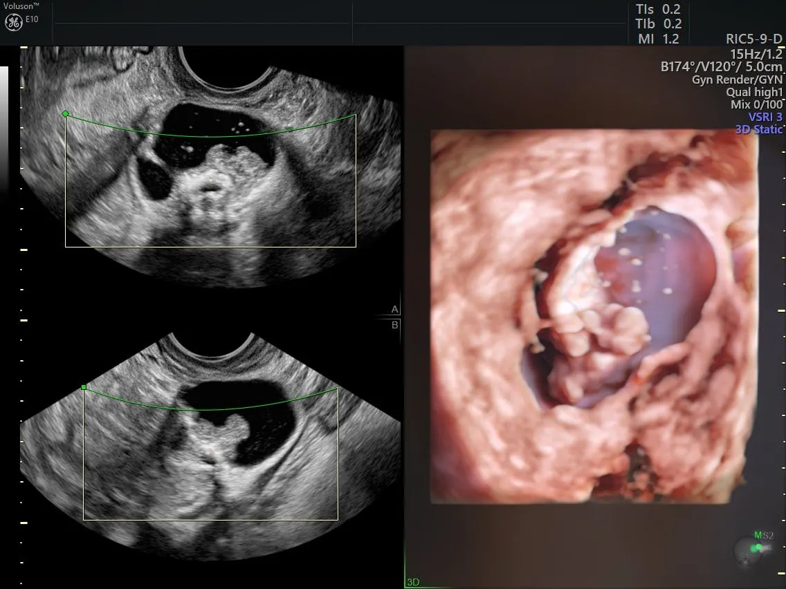
Ultrasound of Adnexal mass
Ultrasound of complex adnexal mass
Why Early Diagnosis of PID Matters
Untreated pelvic inflammatory disease may result in serious complications, such as damage to fallopian tubes or ovarian vein thrombophlebitis. Early detection of pelvic inflammatory disease can eliminate some of the differential diagnosis and ensure the delivery of prompt and comprehensive care.
Gynecologists should use their best judgment when evaluating patients for this condition. Information gathered from imaging tests like transvaginal ultrasound may point to specific causative factors or may help rule out other conditions resulting in similar symptoms, such as endometriosis or appendicitis. In all cases, follow-up testing with CT or MRI may be appropriate based on initial ultrasound findings.

