
Endometrial carcinoma is the fourth most common type of cancer among women in the United States. According to the American Cancer Society, as many as 61,880 new cases of endometrial cancer will be diagnosed in 2019. Endometrial hyperplasia, or excessive thickening of the endometrium, is a known risk factor for endometrial cancer.
Since so many women are at risk for developing this form of cancer, it's crucial for gynecologists to closely monitor — and accurately diagnose — endometrial hyperplasia. Prompt diagnosis and treatment of the condition can reduce the likelihood that it will progress to endometrial carcinoma.
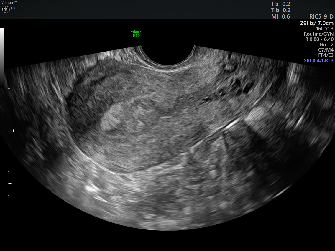
2D image irregular endometrium
The Link Between Endometrial Hyperplasia and Endometrial Carcinoma
Endometrial hyperplasia is typically classified according to cellular characteristics and the presence (or absence) of atypia. The American College of Obstetricians and Gynecologists (ACOG) explains that the standard classification system includes two broad categories: simple hyperplasia, in which the endometrium exhibits benign proliferation of endometrial glands; and complex hyperplasia, in which the endometrium is noticeably irregular, abnormal vasculature is present and cells may be crowded.
The presence of abnormal cells is used to further categorize endometrial hyperplasia. A patient may be diagnosed with simple atypical hyperplasia or complex atypical hyperplasia. The American Cancer Society notes that about 8 percent of women diagnosed with simple atypical hyperplasia develop cancer. Women with complex atypical hyperplasia have the highest endometrial carcinoma risk at around 29 percent.
According to American Family Physician, endometrial cancer is categorized into two types. Type I is more common, accounting for more than 70 percent of endometrial cancer cases. Type I tumors are usually low-grade and generally have a better prognosis compared to Type II tumors. Type II tumors are more likely to be high-grade. In general, Type II tumors have a poorer prognosis and are more likely to metastasize beyond the uterus.
Risk Factors for Endometrial Hyperplasia
Hyperplasia results from unopposed estrogen, which may occur due to hormonal changes, estrogen replacement therapy or certain medications used to treat other cancers. While it's possible for any woman to develop endometrial hyperplasia, the condition most often occurs after menopause.
According to ACOG, other risk factors include:
- Age (older than 35 years).
- Comorbid conditions such as diabetes or polycystic ovarian syndrome (PCOS).
- Early age at menarche.
- Family history of colon, ovarian or uterine cancers.
- Nulliparity.
- Obesity.
- Tobacco use.
In almost all cases of endometrial hyperplasia, patients experience abnormal uterine bleeding. Since bleeding may indicate a variety of gynecological issues, it's important to use the right tools to obtain an accurate diagnosis.
Detecting Endometrial Hyperplasia and Endometrial Carcinoma With Ultrasound
Transvaginal ultrasonography is usually the first diagnostic procedure for suspected endometrial hyperplasia. This test is highly sensitive and is usually much more cost-effective compared to other diagnostic options. The American College of Radiology recommends that postmenopausal women with an endometrial thickness of greater than 5 mm on ultrasound receive further evaluation for the presence of endometrial carcinoma.
Endometrial biopsy is the diagnostic procedure of choice for suspected endometrial cancer, although dilation and curettage and hysteroscopy are also valid diagnostic options. However, these procedures are much more invasive than ultrasound.
Ultrasound may also be used prior to invasive diagnostic tests to identify areas of suspected endometrial carcinoma. 3D ultrasound, which offers a view of the coronal plane, can help determine which areas of endometrial tissue require sampling. A positive tissue sample offers definitive proof of endometrial cancer.
Through advanced technologies like 3D ultrasound, you can distinguish whether abnormal uterine bleeding points to endometrial hyperplasia, investigate other warning signs of endometrial carcinoma and potentially improve your patients' prognosis.
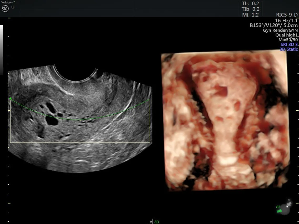
3D image uterine pathology